The next-generation dynamic image analysis tool, FlowCam Nano, is capable of measuring and imaging submicron particles in real time. With its sub-visible particle analysis, FlowCam Nano can detect objects between two micrometers and 300 nanometers in size—the smallest of which is detected with light microscopy.
Users can use FlowCam Nano for the early detection and monitoring of aggregates and contaminants in protein formulations, nanodrug delivery systems, bacterial characterization, material characterization, and bioprocess monitoring.
Image Credit: Yokogawa Fluid Imaging Technologies, Inc.
Use FlowCam Nano for
- Formulation development
- Bioprocessing
- Biomedical research
- Bacterial contamination detection
- Gene therapy aggregate monitoring
- Materials characterization
FlowCam Nano benefits
- High-quality images to identify and detect submicron particle types for product development and quality control
- Instrument setup and data collection are quick and easy, thanks to the user-friendly interface and the autofocus technology
- Rapid detection of API aggregation, as well as other degradation modes that may cause large subvisible and visible particles
- Determine the size and morphology of particles in the 300 nm to 2 µm range that cannot be detected by traditional flow imaging microscopy
- VisualSpreadsheet® software that can visualize, classify, and characterize nanoparticles in real-time
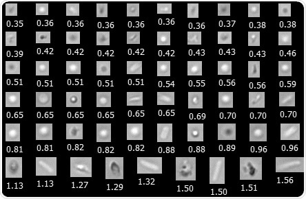
FlowCam Nano captures submicron images of protein aggregates, silicone oil droplets, sucrose aggregates, and E. coli cells. The particle diameter is shown in µm. Image Credit: Yokogawa Fluid Imaging Technologies, Inc.
How it works
Similar to flow imaging microscopy (FIM) instruments, FlowCam Nano consists of a light microscopy apparatus (a stroboscopic LED light source, an objective lens, and a high-speed camera) with a flow cell placed in the path of the light source and objective lens.
Particle analysis is carried out by injecting a sample with a syringe pump. Particle images passing through the optics are captured and recorded automatically. After analyzing the microscopic images, the size, morphology, concentration, and types of particles in the sample can be determined.
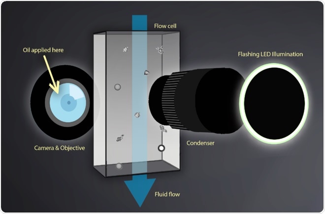
Image Credit: Yokogawa Fluid Imaging Technologies, Inc.
A high-magnification (40X) objective lens and a thin layer of immersion oil reduce the numerical aperture between the flow cell, the objective, and the condenser of FlowCam Nano, providing higher resolution than standard flow imaging microscopy.
The patented FlowCam Nanotechnology magnifies particles near and below the size limit of conventional FIM instruments, extending the range of particle detection and imaging capability to submicron particles.
FlowCam Nano specifications
- Detects particles in the range of 300 nm to two µm
- Magnification: 2X, 4X, 10X, 20X
- Minimum sample volume of 100 μL
- Frame rate: up to 130 frames per second
- Flow cell: 60 µm depth
- Sample processing capability: 20 µL/minute
- Camera resolution of 1920 x 1200 pixels, available in color and monochrome
- Autofocus capability
- The system offers dual-channel fluorescence detection with excitation options at 488 nm, 532 nm, and 633 nm.
- Automated self-cleaning and self-rinsing cycles
- Compatible with the Automated Liquid Handler (ALH for FlowCam)
- Equipped with the image analysis software VisualSpreadsheet®
- Optional modules include VisualAI™ for identifying protein aggregates/silicone oil droplets and a compliance package to ensure standards are met, such as 21 CFR Part 11
Explore FlowCam applications
- Biopharma and pharma
- Water quality monitoring
- Plankton research
- Harmful algal blooms
- Food and beverage
- Industry