Versatile optical imaging by MILabs
Video credit: MILabs
Features
- Versatile ultra-high-sensitivity imaging system for standalone benchtop optical imaging
- High-throughput: ability to image up to 10 animals simultaneously on a standalone OI system
- Broad wavelength selection for bioluminescence, fluorescence, and Cherenkov applications
- Interchangeable with in-line CT for OI/CT tomographic functionalities of deep tissues & organs
- The OI/CT configuration delivers quantitative 3D/4D optical tomography results for bioluminescence and fluorescence applications, including imaging of deep tissues
- Upgradeable with PET and/or SPECT modalities
Versatile in-vivo optical imaging
MILabs offers ideal and powerful optical imaging solutions for a vast variety of preclinical applications ranging from molecular and functional imaging to translational research. By using the latest state-of-the-art components, the benchtop U-OI system blends high performance with affordability and industry-leading upgradability. The benchtop 2D imaging unit offers imaging of up to ten mice simultaneously with a broad choice of optical filters, making it optimally configurable for bioluminescence, fluorescence, and Cherenkov imaging. The system can be also tailored to users’ applications from the default large-area ultra-sensitive 1 MP (HR) to optional 4 MP (UHR) cameras.
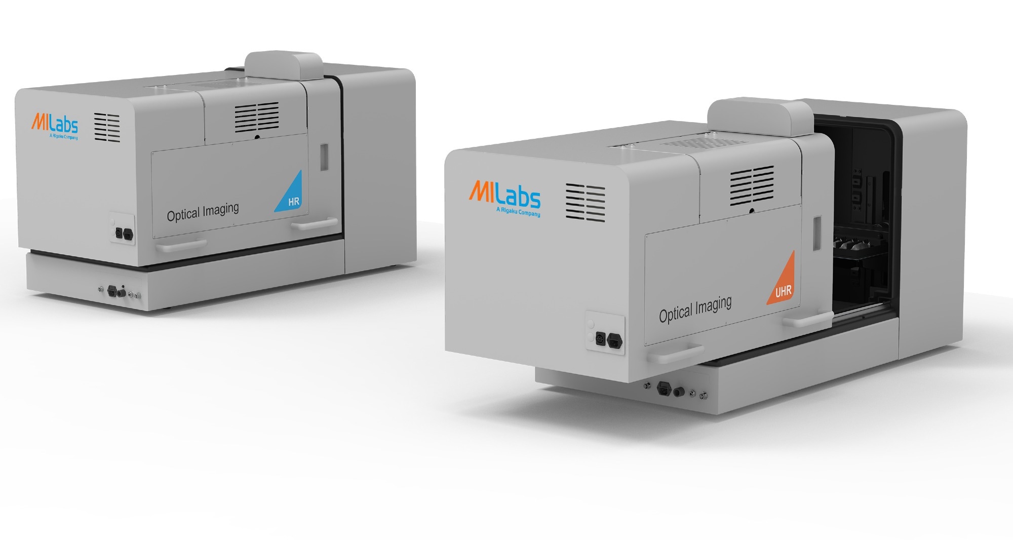 Image credit: MILabs
Upgradeable to integrated OI/CT and unique 3D/4D CT-guided optical tomography
Additionally, the U-OI system can be seamlessly integrated with MILabs’ CT system by simply transferring the optical module from the benchtop docking station to the in-line X-ray CT. The integrated OI/CT allows for optical and CT imaging in parallel for dedicated multimodal applications. This allows you to switch freely between in-line and benchtop (or off-line; standalone) modes: the in-line mode enables OI/CT acquisitions on a single-floor standing platform, while the separate benchtop mode is still available.
The seamless in-line mode allows you to perform optical tomography with the help of MILabs’ unique bioluminescence tomographic ("BLT") and/or fluorescence tomographic ("FLT") imaging functionalities. Segmented CT images from the same animal(s) are used to guide 3D/4D BLT and/or FLT images for more accurate reconstruction and quantification of deep tissues and dark organs.
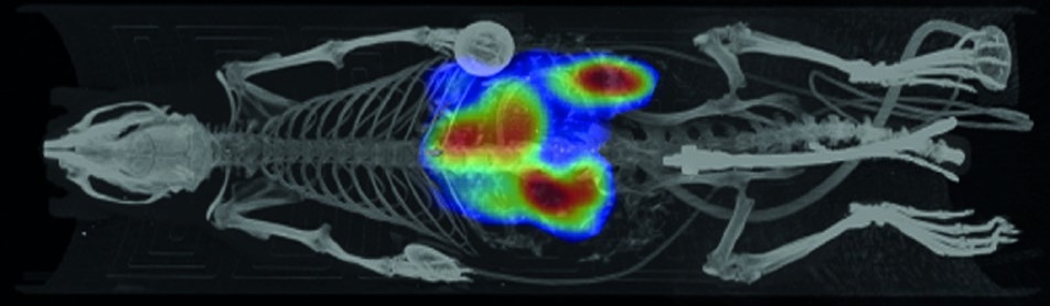
Figure 1: Anatomical co-registration of 2D optical and 3D CT using Perspective MIP. Image credit: MILabs
Upgrade with PET/SPECT
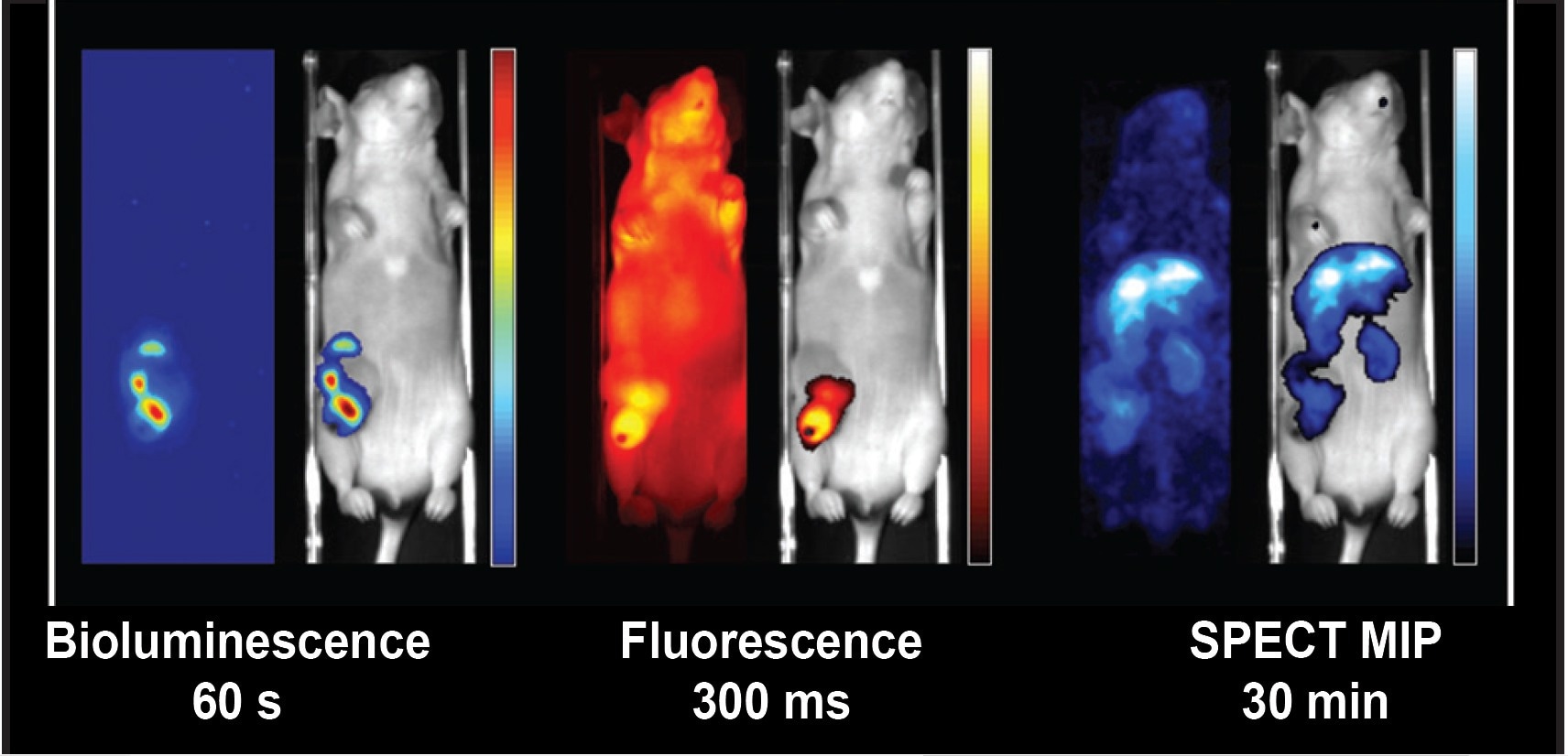 Figure 2. Example of complementary registered bioluminescent, fluorescent, and nuclear images in one scanning session with a single dose of anesthesia [Multimodality Molecular Imaging of Lung Disease Using PET, CT and Optical Imaging, SSG07-09 Oral Presentation, RSNA 2019].
Translation of optical imaging results to clinically compatible PET or SPECT data is easily accomplished by adding the desired nuclear modality to the OI/CT system. MILabs can offer any combination of in-line PET, SPECT, optical, and CT on a single platform. These modalities can be added at any time to suit your requirements and budget. This makes one-to-one translation from basic optical research data to clinically compatible nuclear imaging technologies possible and avoids animal shuttling with inconsistent anesthesia conditions and image registration challenges.