The Oxford Instruments witec360 sets a new standard in Raman imaging and correlative microscopy, delivering nanoscale analysis capabilities to meet the evolving needs of researchers in both academic and industrial settings.
The witec360, the successor to the pioneering alpha300 series, establishes new benchmarks for performance and broadband capability. Its inherently modular design offers both immediate and enduring value, facilitating straightforward adoption for those new to Raman spectroscopy while providing the adaptability required to accommodate diverse and rapidly advancing applications.
Benefits
Acquire research-grade results
The system's fully optimised optics ensure best-in-class speed, sensitivity, and resolution.
Experience unprecedented versatility
Broadband capabilities offer the spectral flexibility required for challenging research tasks and diverse applications in multi-user facilities.
Gain comprehensive insight
The seamless integration of imaging techniques enables the correlation of chemical and structural properties, resulting in a more comprehensive understanding of samples.
Configure the microscope for current and evolving experiments
Modular design provides extensive capabilities and upgrade options, enabling tailored solutions and scalability to match individual requirements and resources.
Enhance productivity with intelligent automation
Advanced hardware automation and intuitive software streamline data acquisition and analysis, leading to enhanced operational efficiency and rapid user onboarding for individuals with varying levels of experience.
Ensure consistency and transparency
Multi-user management, internal calibration, automated reporting, and streamlined analysis collectively promote reproducibility and support adherence to both institutional and regulatory standards.
Advanced confocal Raman technology for every task
- Time-resolved measurements for monitoring reactions and material changes
- Point-by-point spectral acquisition for accurate measurement at individual locations
- Line scans to facilitate depth profiling across predetermined axes
- 2D area scans for high-resolution imaging and quick overview mapping
- 3D analyses that reveal internal structures layer by layer
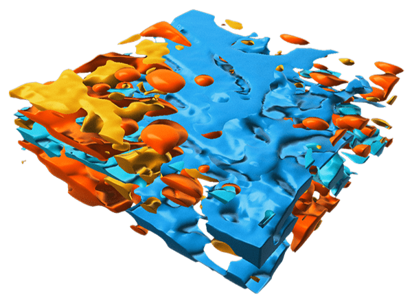
3D Raman image of a cosmetic emulsion rendered in Imaris. Dimensions: 30 x 30 x 10 µm3. Image credit: Oxford Instruments
Modularity
A system designed to meet the needs
Flexible – sustainable – future-proof
The witec360 system is built upon a modular architecture, allowing for the precise customisation of configurations to address diverse experimental challenges. Its offerings encompass a spectrum of solutions, from fixed setups optimised for single applications to specialised instrumentation for advanced academic research, and tools engineered to satisfy industry benchmarks and multi-user operational demands. The design's inherent flexibility ensures the witec360's capacity for evolution and expansion in response to future requirements.
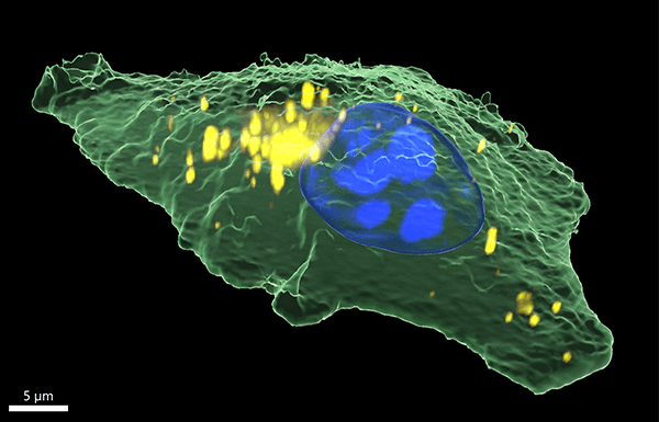
3D Raman image of a human epithelial cancer cell rendered in Imaris. Sample courtesy of Dr. Irina Estrela-Lopis and Tom Venus, Institute of Medical Physics and Biophysics, Leipzig University, Germany. Image Credit: Oxford Instruments
Explore the options within the witec360 ecosystem
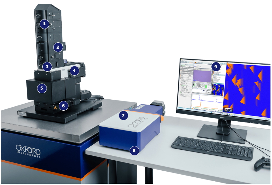
Image Credit: Oxford Instruments
1. Modular input and output couplers
- Individual combination of imaging modes and correlative techniques
- Polarisation-maintaining optics
- Optimal alignment for an ideal beam path and high throughput
- RayShield coupler and RayLine filters for low-frequency signal detection
- Manual or automated operation
- A polarisation unit that enables the free rotation of the polarisation angle or the creation of light that is circularly polarised.
2. Lasers
- Fiber-coupled lasers for the highest flexibility and efficiency
- Wide selection of available wavelengths and power levels
- Six wavelengths can be integrated into a single setup
- Highly accurate fiber couplers allow for rapid switching between laser sources
- Absolute laser intensity control via patented TruePower™ technology
- Automatic shutter control
- Optical components that maintain polarisation
3. Illumination and detection setup
Adaptable lighting and sensing capabilities suitable for diverse areas of study and various sample shapes.
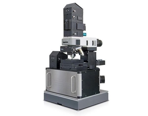
- Upright or inverted microscope body for illumination from above or below
- Supports both transmitted and reflected light modes
- BeamFlex functionality provides maximum adaptability for upright and inverted imaging, covering reflection and transmission settings.
4. Whitelight/widefield microscopy
- Class-leading Köhler illumination ensures even, high-contrast imaging throughout the full field of view.
- Premium white-light microscopy delivers high-quality images across multiple imaging modes, including Brightfield, Darkfield, and DIC.
- Supports operation in both reflection and transmission modes.
- Optional polarisation configuration available
- Fluorescence microscopy can be optionally integrated to enable multi-modal imaging.
5. Objective turret
- Holds up to six objectives at the same time.
- Works with high-performance objectives from well-known manufacturers.
- Offers an optional software-controlled turret rotation feature to streamline workflows.
6. Sample positioning and handling
Sample positioning
- For accurate scanning in the X, Y, and Z axes, both manual and motorised stages are available.
- Piezo stages are available for applications requiring the highest level of spatial resolution and accuracy.
- Standard travel ranges are available, from 25 mm × 25 mm up to 350 mm × 300 mm.
Sample handling accessories
- Specific sample holders accommodate diverse instruments and sample shapes.
- Environmental control units, including heating and cooling platforms and stage-top incubators, are available.
- These stages facilitate specialised experimental conditions.
7. Spectrometers
Hexalight
- The spectrometer's lens-based, mirror-free architecture guarantees exceptional sensitivity and throughput.
- With up to six gratings installed, the device provides both maximum resolution and versatile application, eliminating the need for manual adjustments.
- The integrated design allows for easy switching between high-resolution and broadband acquisition of Raman and PL spectra.
- The unique Olaf-Hollricher-Design (OHD) lenses offer genuine broadband performance across the 350 to 1100 nm range.
- Automated grating selection and precise positioning are made possible by an innovative harmonic drive.
- Users can choose between 300 mm and 600 mm focal length options.
Monolight
- Suited for particular uses, this solution employs a single, fixed grating.
- Available configurations cater to lasers operating across a spectrum from ultraviolet (266 nm) to near-infrared (1064 nm).
Detectors
- Oxford Instruments Andor cameras are highly sensitive and designed for advanced Raman imaging.
- High quantum efficiencies
- Rapid acquisition times
- Enhanced imaging quality with fewer artifacts
- Very low dark current
- A range of options, from budget-friendly to high-end (Front Illuminated, Back Illuminated, Electron Multiplying, Open Electrode)
- InGaAs array detectors for expanded capabilities in the NIR excitation range
8. Compact footprint
- Adaptable arrangement of all elements
- Minimised thermal and vibration disruptions during measurement
- Suitable for use inside a glove box or enclosed environment
9. Software
- Software suite for data acquisition and analysis
- Adaptable software configurations through a modular architecture
- User-interface optimised for effective instrument control
- Smooth control of supported correlative techniques
- Smart data analysis and visualisation
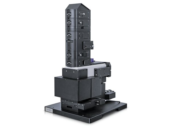
Upright. Image Credit: Oxford Instruments
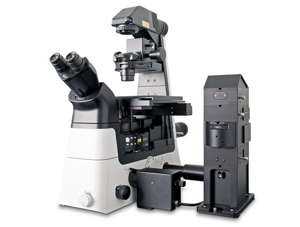
Inverted. Image Credit: Oxford Instruments
BeamFlex. Image Credit: Oxford Instruments
Advanced software packages
ParticleScout: Rapid and straightforward Raman particle identification and classification
TrueMatch™: Raman spectral database management software for streamlined compound identification
Workflow Manager: Enables automated and recurring measurement workflows
Multi-user Management: Data access and instrument function control for individual users and groups
Programming Interface: Customised measurement procedures for specific applications
DaVinci: Precise nanomanipulation and nanolithography tool
The path to the Raman microscope
- Choose the microscope basis (upright/inverted)
- Choose the microscope parts
- Include sophisticated imaging and analysis choices
- Personalise with accessories
Correlative microscopy
Comprehensive sample analysis with Raman and correlative techniques
The witec360 platform integrates correlative methodologies, thereby extending its analytical capacity to yield comprehensive insights into chemical, topographic, mechanical, and structural characteristics at a unified sample location. The associated software provides streamlined operational management, facilitates automated data correlation, and enables intuitive image superimposition for optimised analytical workflows.
Upgrade options for correlative imaging techniques are available for both current witec360 systems and legacy alpha300 series microscopes.
Correlative imaging techniques
Scanning electron microscopy (SEM)
Critical for material analysis, nanotechnology, and geosciences, SEM techniques yield vital nano-structural, crystallographic, and elemental insights.
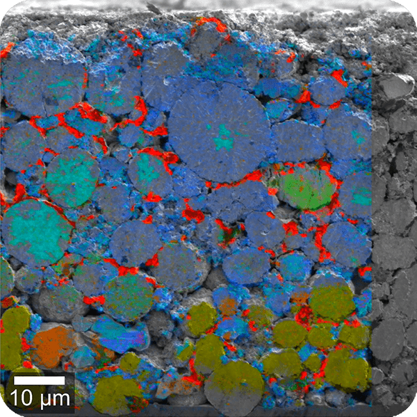
Image Credit: Oxford Instruments
Photoluminescence (PL) imaging
PL is a widely employed technique for material science and semiconductor characterisation, delivering critical insights into optoelectronic properties.
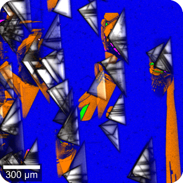
Image Credit: Oxford Instruments
Atomic force microscopy (AFM)
AFM offers crucial insights into nanoscale surface properties (topography, adhesion, and stiffness) for materials science and nanotechnology applications.
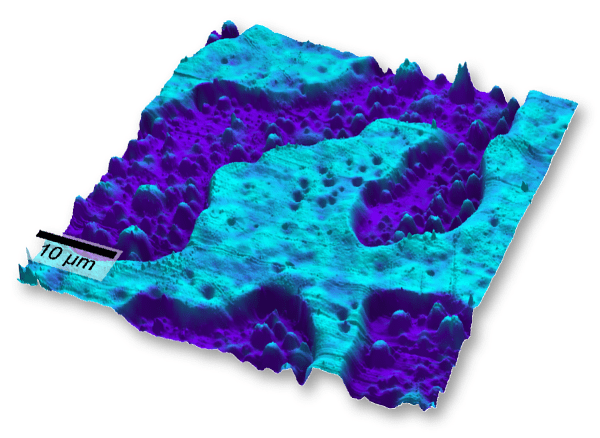
Image Credit: Oxford Instruments
Topographic Raman imaging
TrueSurface facilitates simultaneous topographic and Raman measurements on samples exhibiting irregular surfaces, including rocks, tablets, or micro-structured materials.
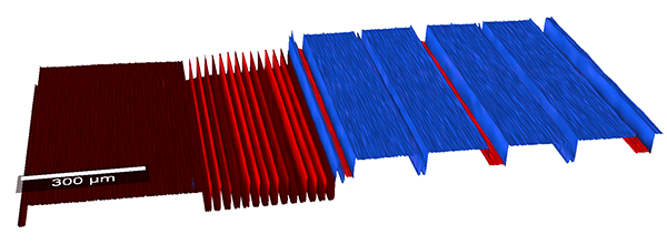
Image Credit: Oxford Instruments
White-light microscopy options
Köhler illumination in witec360 microscopes ensures optimal white-light images for efficient region of interest location and visualization.
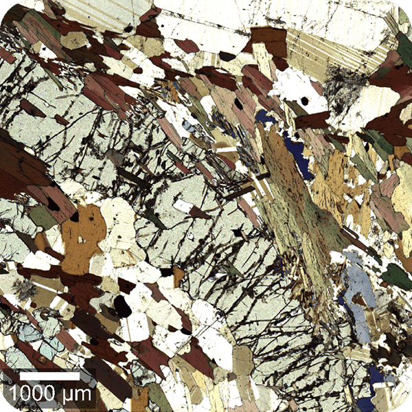
Image Credit: Oxford Instruments
Scanning near-field microscopy (SNOM)
SNOM accomplishes optical imaging with resolutions down to ~60 nm, ideal for analyzing nanomaterials and complex samples.
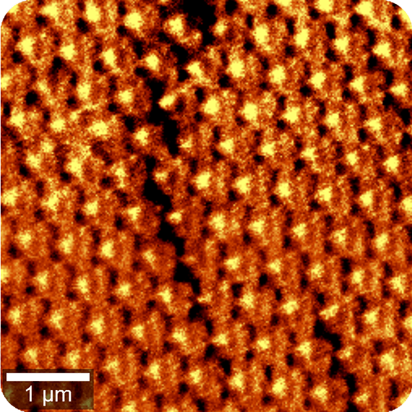
Image Credit: Oxford Instruments
Fluorescence microscopy
Raman data can be combined with fluorescence microscopy for life science applications that use fluorescent labeling.
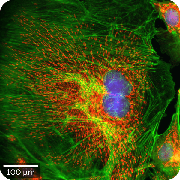
Image Credit: Oxford Instruments
Time-correlated single photon counting
TCSPC techniques, including TLM and FLIM, are instrumental across semiconductor, optoelectronics, and life sciences research domains.
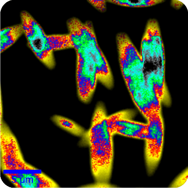
Image Credit: Oxford Instruments
Antibunching measurements
For quantum computing and cryptography, single-photon emitters (SPEs) are identified using correlative Raman, Photoluminescence (PL), and antibunching techniques.
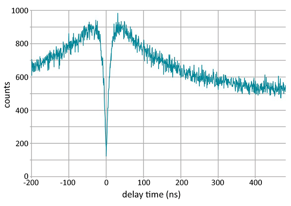
Image Credit: Oxford Instruments
Second harmonic generation (SHG)
SHG is highly responsive to non-centrosymmetric structures. It is well-suited for examining the characteristics of crystals in 2D materials and providing label-free observations in the life sciences.
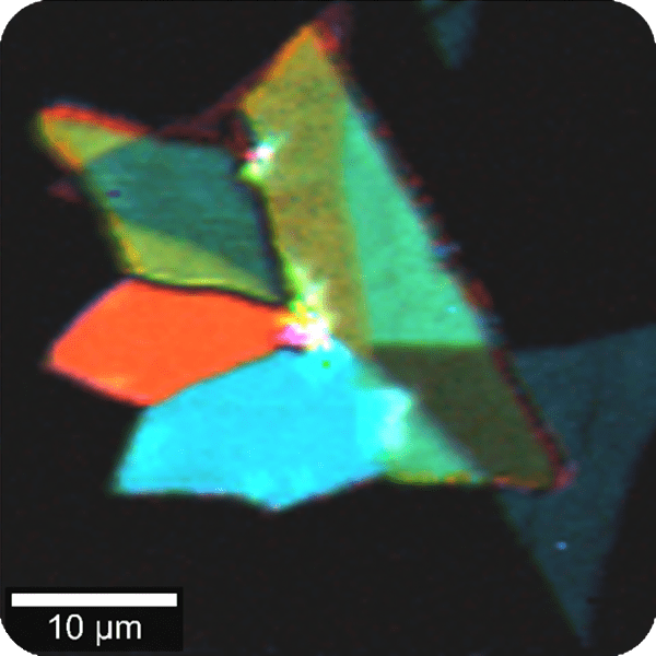
Image Credit: Oxford Instruments
Performance
Class-leading performance
The witec360 establishes the benchmark for performance in confocal Raman imaging. Its components are meticulously optimised for superior transmission efficiency and consistency, ensuring rapid and precise data acquisition. This design significantly streamlines imaging workflows and enables the successful execution of even the most complex applications.
- Maximised signal intensity for the broadband range
- 350 - 1100 nm within the same core system
- Extended options from UV (266 nm) to NIR (1064 nm)
- Diffraction-limited spatial resolution
- Depth resolution < 950 nm (dependent on excitation wavelength)
- Lateral resolution < 300 nm (dependent on excitation wavelength)
- Exceptional spectral resolution
- Resolution is achieved down to 0.1 cm-1 relative wavenumbers (at 633 nm excitation)
- Ultra-fast Raman imaging
- Acquisition speeds are below 1 ms per spectrum without compromising resolution
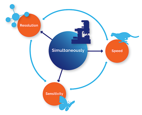
Image Credit: Oxford Instruments
Hexalight - the core of witec360’s performance
The witec360 incorporates the innovative Hexalight spectrometer. This spectrometer is equipped with lens-based, mirror-free optics and 6 grating positions, ensuring optimal throughput and spectral flexibility across the broadest wavelength range.
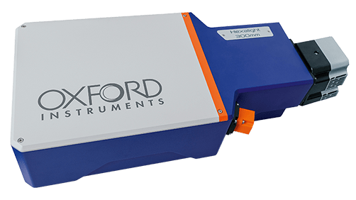
Image Credit: Oxford Instruments
Automation
Effortless precision with advanced automation
Advanced automation options for witec360 microscopes maximise efficiency and simplify workflows. These options help ensure consistency and deliver high-quality results that users can rely on.
Key benefits:
- Ease of use: Simplifies complex tasks and enables advanced measurements, catering to both novice and expert Raman imaging users.
- Streamlined workflow: Automated adjustment procedures enhance efficiency, dedicating more time to research.
- Enhanced reproducibility: Ensures high-quality, reliable results through automated handling of repetitive tasks and hands-free optics positioning.
- Remote operation: Facilitates complete remote control of measurements and microscope operation, suitable for enclosed environments such as glove boxes.
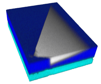
3D Raman image of a defect in a SiC wafer. Dimensions: 180 x 245 x 7 µm3. Image Credit: Oxford Instruments
Automation for every user
Multi-user labs:
This solution accommodates varying experience levels, streamlining workflows with easy-to-use shared equipment.
Industry researchers:
It handles recurring experimental tasks quickly and precisely to accelerate development
Raman newcomers:
Advanced tasks are simplified, enabling confident operation with intuitive onboarding.
Raman experts:
Push the boundaries with advanced automation for complex experiments.
witec360 automation key features
Instrument setup and imaging control
| Feature |
Benefit |
|
AutoBeam – One click precision alignment
This system offers automatic laser beam path alignment, employing a certified reference sample to achieve diffraction-limited resolution and stable performance. It incorporates a memory function for various laser alignments and supports full remote operation.
|
It is optimally designed for multi-user laboratories and enclosed setups, providing hands-free, repeatable alignment for consistent, high-quality outcomes.
|
|
TrueCal – Calibration made easy
Automated, pre-configured calibration routines are included, ensuring the system remains spectrally aligned and prepared for experiments.
|
This makes it an ideal solution for repetitive or regulated workflows demanding precision, compliance, and quantitatively accurate results.
|
| Feature |
Benefit |
|
TrueComfort – Simplify everyday operations
Enhanced convenience and accelerated imaging through streamlining essential operations:
- Motorised Köhler illumination for optimal light settings
- Quick switching between bright-field and Raman modes
- Software-controlled objective revolver for one-click rotation
|
An economical solution designed to enhance efficiency and simplify routine imaging operations.
|
|
TrueSurface – Always in focus
Actively maintains perfect focus during the entire measurement for sharp and high-quality images.
|
Optimized for large-area scanning and accommodating samples with complex geometries, including curved, tilted, and structured surfaces.
|
|
Motorised laser coupler – Expand the wavelength options
Features software-controlled selection from up to six laser wavelengths, covering the UV to NIR spectrum.
|
Designed for multi-wavelength experiments and a broad range of samples necessitating varied excitation conditions.
|
|
TruePower™ – Absolute laser power control
Absolute laser power is precisely controlled throughout measurements, ensuring consistent and reproducible illumination with an accuracy better than 0.1 mW (extending to 0.1 µW in advanced configurations).
|
This functionality is indispensable for energy-dependent experiments and for sensitive samples requiring stringent laser power regulation.
|
|
Motorised polarisation modules – Versatile and accurate
Automated adjustment of freely rotatable excitation polarisers and detection analysers simplifies and accelerates polarisation-sensitive and polarisation-resolved series measurements.
|
Enhance the research into molecular orientation and anisotropic materials through increased usability and accelerated workflows.
|
|
Motorised and piezo-driven stages – Precision made easy
Achieve precise, automated sample placement using integrated motorized and piezo-driven scan stages.
|
This capability is crucial for comprehensive large-area mapping, automated focus on specific regions, and accurate re-examination of prior measurement points
|
Data processing
Leverage the sophisticated software to simplify data processing and analysis, enabling rapid result interpretation.
| Feature |
Benefit |
|
Software Suite Analysis Wizard
Simplifies data analysis with guided, click-through workflows.
|
Training time is reduced, and data analysis is accelerated for users across all experience levels.
|
|
TrueComponent analysis
Sample components within an image are automatically identified and differentiated.
|
Complex samples undergo fast and comprehensive analysis, with spectral unmixing made easy.
|
|
TrueMatch
Measured spectra are quickly matched to known molecules through the use of renowned spectral databases.
|
Direct peak assignment is achieved without interpretation or mathematical evaluation.
|