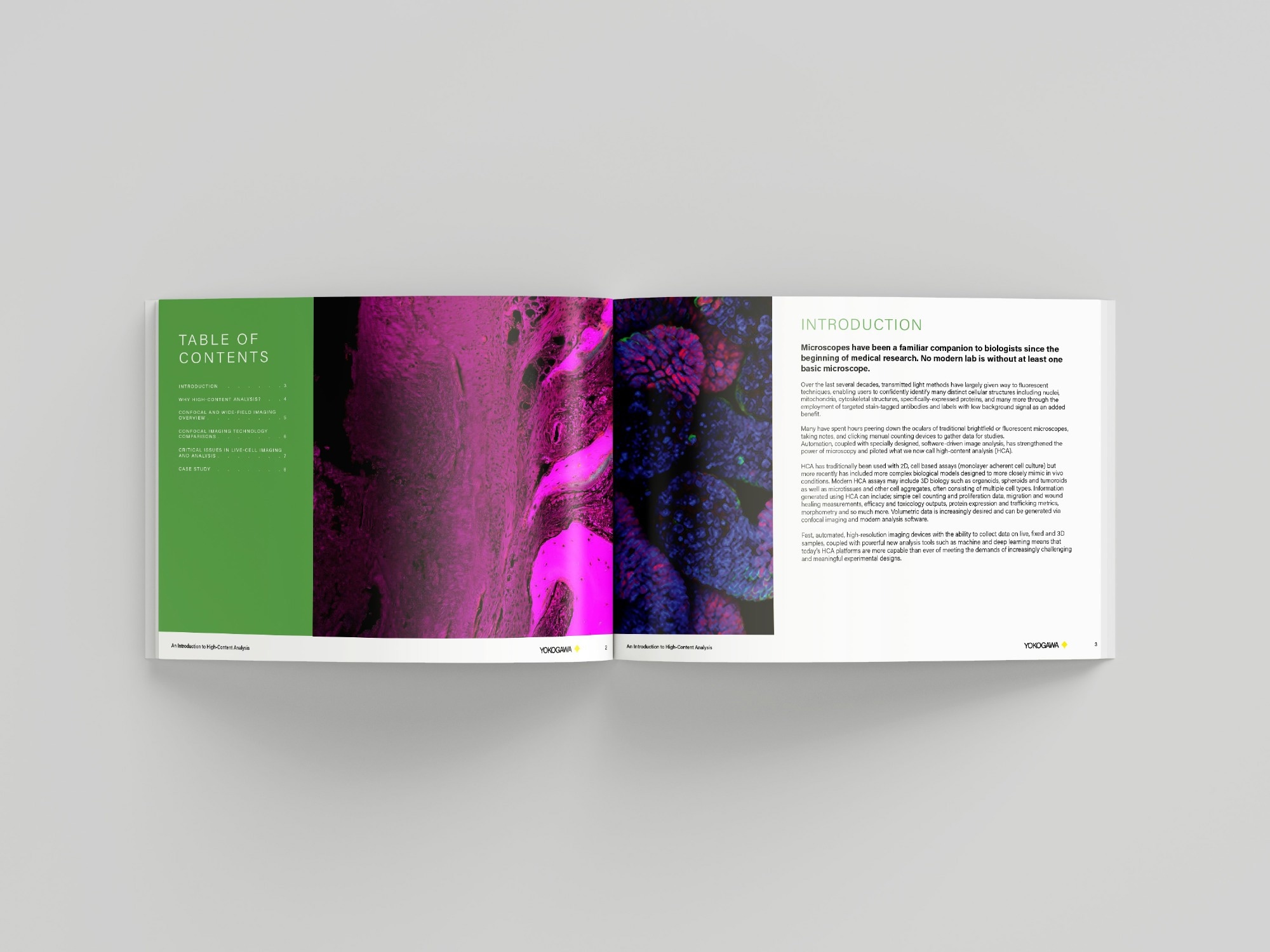
Microscopes have been a constant companion for biologists since the inception of medical research. There is no modern lab without at least one basic microscope.
Fluorescent techniques have largely replaced transmitted light methods in recent decades, allowing users to confidently identify many distinct cellular structures, such as nuclei, mitochondria, cytoskeletal structures, and specifically expressed proteins, among others, by employing targeted stain-tagged antibodies and labels, with a low background signal as an added benefit.
Many scientists have spent hours staring under the oculars of conventional brightfield or fluorescent microscopes, scribbling notes, and clicking manual counting mechanisms to collect data for research.
Automation, combined with specially designed, software-driven image analysis, has enhanced the capabilities of microscopy and paved the way for high-content analysis (HCA).
The Yokogawa Life Science High-Content Analysis (HCA) eBook provides a concise introduction to high-content imaging and analysis, a powerful tool for modern life science research.
It demonstrates how automated microscopy paired with modern image analysis allows for multiparametric, quantitative assessment of cellular phenotypes on a large scale, pushing beyond single-endpoint experiments.
The eBook discusses the fundamental principles of HCA, major imaging modalities, and the practical aspects of designing robust, reproducible trials.
The eBook demonstrates how HCA is applied in drug development, toxicity testing, disease modeling, and live-cell investigations, utilizing real-world examples and workflow instructions, as well as complex 3D and physiologically relevant models.
It also addresses typical issues such as data quality, phototoxicity, and large-scale image analysis, enabling researchers to develop accurate, high-confidence conclusions from high-content datasets.
About Yokogawa Life Science
We have 30 years of experience in this life science field and will respond to customer's problem solving with cutting-edge solutions.
Our confocal scanner unit CSU series enables 3D observation of the cells in detail and dynamics of organelles inside cells. Since the CSU series is capable of high-speed shooting, it is also suitable for observing high-speed life phenomena. In addition, the CSU series is a multi-point confocal method which is extremely gentle to cells, best suitable for long-term live cell observation.
Sponsored Content Policy: News-Medical.net publishes articles and related content that may be derived from sources where we have existing commercial relationships, provided such content adds value to the core editorial ethos of News-Medical.net, which is to educate and inform site visitors interested in medical research, science, medical devices and treatments.