Shutterstock | Digital Photo
Raman tissue imaging is considered to be a unique method capable of simultaneously describing the molecular composition and the distribution of multiple chemical species in tissues at high spatial resolutions, without labeling.
Raman imaging makes use of a laser to provide a chemical fingerprint at every point of the analyzed area. One can process the data in order to reveal a multitude of information, such as:
Discriminate Diseased and Healthy Tissues
- Distinguish tissue types by their total chemical signatures – fluorescent or colorimetric labeling not needed
- Differentiate healthy and diseased tissues accurately and objectively
- No need for disease marker discovery and targeting
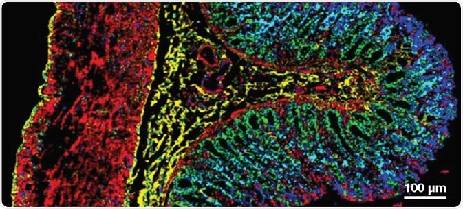
Clearly visualize the tissue organization with Raman images - healthy rat colon crypt.
Demarcate Tissue Regions Chemically
- Visualize anatomical layers and cell types
- Generate detailed chemical images with sub-micrometer spatial resolution
- Reveal important zonal information:
- complexity of the tissue organization
- relative abundance and distribution of chemical species
- boundary and size of diseased area
- tumor infiltration
- Unveil the morphological and chemical changes in varied samples
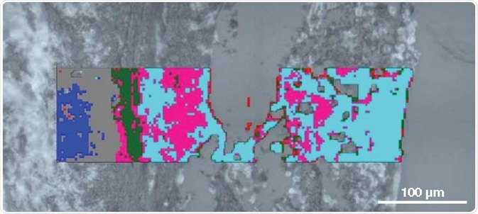
Understand cancer progression - examine melanoma invasion in skin construct.
Potential Pathology Tool
- Discriminate cancer stages with high specificity and sensitivity
- Determine early onset disease markers
- Detect biochemical changes linked with cancer formation and progression
- Provide histological images without labeling
- Define tumor margin
The Perfect Technique for Biological Research
- Study the concentration, conformation, distribution, redox and spin states, and orientation of biomolecules
- Compare these properties between samples
- Obtain valuable insight and improved understanding of the biological system of interest
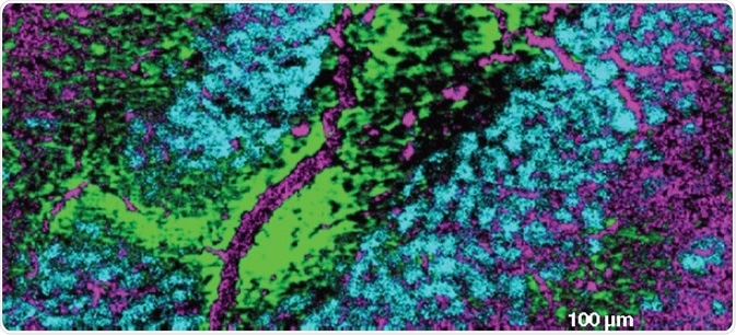
Reveal the distribution of cells and assess their molecular profiles - neurons and glial cells in the rat brain displaying different cytochrome levels and redox states.
inVia. The Perfect Raman Tissue Imaging Tool
- Research-grade Raman microscope
- StreamLine™ imaging technology for high speed mapping without damaging tissues
- Surface option to attain the best images from uneven surfaces
- StreamLine imaging with Slalom for a rapid overview of the tissue samples
- High confocality StreamHR™ imaging to scrutinize minor details
- Flexibility to shift between high and standard confocal imaging
- Queue up measurements to maximize data collection

The Renishaw inVia Raman microscope.

This information has been sourced, reviewed and adapted from materials provided by Renishaw plc.
For more information on this source, please visit Renishaw plc.