The brain is never completely at rest. Even without external input, it produces spontaneous neural activity that creates synchronized fluctuations across different regions - a process known as functional connectivity (FC).
Initially described using resting-state fMRI (Biswal et al., 1995), FC portrays the brain’s intrinsic functional organization, which underlies cognition and behavior and is altered in various neurological and psychiatric conditions. Its evolutionary conservation (Gutierrez-Barragan et al., 2024, Nat Comm), how it aligns with anatomical connectivity (Suarez et al., 2020, TiCS), and how it overlaps with canonical networks (such as sensory, motor, and default mode systems; Smith et al., 2009, PNAS) highlight its key role in neural organization.
FC alterations are involved in conditions from Alzheimer’s (Al-Ezzi et al., 2024, Comm Biol) to schizophrenia (Sheffield and Barch, 2016, Neurosci Biobehav Rev) and autism (Cai et al, 2025, J Biophotonic). They are thus a biomarker and a window into disease mechanisms at the same time.
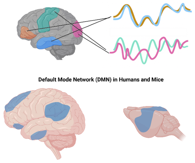
Image Credit: Iconeus
Even though resting-state fMRI is the gold standard for human use, functional ultrasound imaging (fUSI) has emerged as a helpful aid in preclinical research (Osmanski et al., 2014, Nat Comm). Its mix of high sensitivity and fine spatiotemporal resolution enables fUSI to overcome the typical constraints inherent to fMRI in small-animal models, opening new possibilities for the study of FC in both health and disease.
Functional ultrasound imaging: Principles and advantages
fUSI builds upon the principle of neurovascular coupling. Activation of neurons causes local blood vessels to dilate, increasing cerebral blood volume (CBV). Ultrafast Doppler sequences identify these CBV fluctuations with a high degree of sensitivity. The approach differs from traditional Doppler, which works by sequentially scanning tissue. Instead, fUSI captures entire planes at a rate of thousands of frames per second (Mace et al., 2011, Nat Methods).
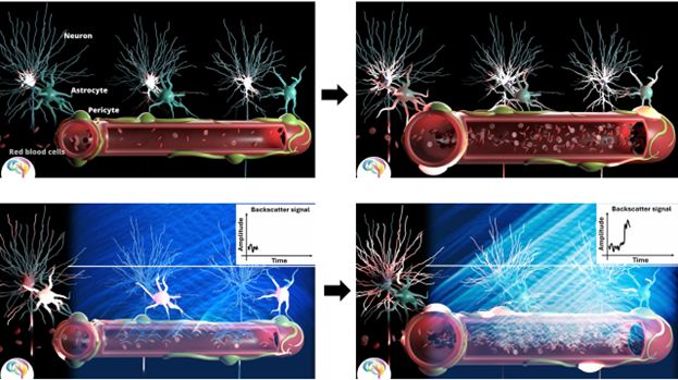
Image Credit: Iconeus
This technique delivers:
- Elevated spatial resolution (100 µm) and temporal resolution (10–400 ms)
- Identification of CBV changes from a minimum of 2% above baseline
- A signal-to-noise ratio that can map fine-scale vascular dynamics
Such properties make fUSI an ideal fit for studying spontaneous activity and inter-regional correlations that characterize FC in preclinical use cases.
Use in functional connectivity research
Functional ultrasound imaging (fUSI) has revolutionized knowledge of large-scale brain networks, delivering special insights into their organization, plasticity, and dysfunction alike. Research in mice has demonstrated strong similarities with human neuroimaging, such as the existence of a default mode network (DMN) whose patterns of connectivity change according to brain state (Ferrier et al., 2020, PNAS).
Such an evolutionary conservation validates fUSI as a translational tool and highlights its ability to record dynamic network conditions, from resting states to sensory processing.
Aside from mapping resting-state activity, fUSI has provided insight into the neural mechanisms that underpin behavior. For instance, in learning and memory tasks, it has been able to trace the reorganization of connectivity among the amygdala, hippocampus, and prefrontal cortex - changes that reflect behavior like fear responses (Grohs-Metz et al., 2022, Behav Brain Res). By helping to visualize how networks become stronger or weaker over time, fUSI delivers insight into how memory consolidation and adaptive plasticity work.
The technology’s sensitivity also applies to disease models, where it has revealed disrupted connectivity in neurological conditions. Among models of peripheral neuropathy (Anzibar Fialho et al., 2024, Sci Rep) and schizophrenia (Zhou et al., 2024, biorXiv), fUSI has been able to link circuit-level dysfunction to behavioral symptoms, and thus bridge the gap between molecular pathology and systemic brain activity. These findings position fUSI as a powerful instrument for understanding the mechanisms underlying disease and coming to know biomarkers for neural dysfunction.
In pharmacology, fUSI has shown potential for accelerating drug development by uncovering how substances such as opioids influence brain-wide connectivity. These “network fingerprints” are linked to therapeutic effects (e.g., analgesia) while setting them apart from side effects, providing a nuanced technique for assessing both drug action and safety (Mariani et al., 2024, biorXiv).
These use cases underscore fUSI’s ability to propel fundamental neuroscience and clinical research spanning evolutionary comparisons and behavioral neuroscience to therapeutic innovation.
Iconeus solutions for functional connectivity research
High-quality FC research requires more than sensitivity alone; it demands integrated tools for acquisition, analysis, and reproducibility alike. Iconeus offers a comprehensive workflow that has been customized for preclinical FC studies:
Hardware
- Iconeus One system: This portable imaging station features plane-wave ultrasound technology and an integrated, robust workstation. A height-adjustable keyboard and monitor allow users to conduct experiments comfortably. The system also includes a four-axis motorized scanning platform ideal for imaging head-fixed animals.
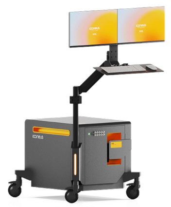
Image Credit: Iconeus
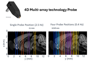
Image Credit: Iconeus
- Multi-array probe: It uses four planes simultaneously, allowing for whole-brain mouse coverage in 2.4 seconds. It is usable transcranially and with craniotomy.
- RCA probe: Row-column addressed technology facilitates volumetric imaging of an 8 × 8 x 20 mm brain volume in 400 ms.
Software
- IcoScan: A user-friendly acquisition platform that has been modified for fUSI tests. Extremely easy to customize and easy to use.
- IcoStudio: Sophisticated analysis environment supporting FC-specific workflows such as correlation matrices, seed-based connectivity maps, flexible time-windowing, denoising, and signal exploration.
- IcoLab: Batch and group-level processing instrument for reproducibility. It combines filters with analysis pipelines, rejects artifacts, and creates summary reports (PowerPoint) and exportable data files for more statistical analysis.
These tools, taken together, offer a smooth transition from acquisition to publication-ready outcomes, making fUSI-based FC research simultaneously powerful and accessible.
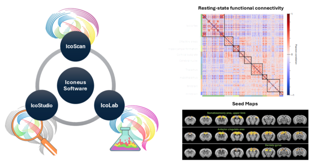
Image Credit: Iconeus
Conclusion
Functional connectivity enables new insights into how the brain is organized and dysfunctional. With its unique blend of sensitivity, spatial and temporal resolution, and compatibility with preclinical models, functional ultrasound imaging is quickly becoming a method of choice for studies involving FC.
Integrating advanced probes with a dedicated software suite, Iconeus offers a comprehensive solution for scientists seeking to explore functional connectivity - spanning the fundamentals of neuroscience and systems-level research to disease models and pharmacology.
Want to continue learning? Watch this webinar on the topic
About Iconeus
Iconeus is a Paris-based company, founded by the inventors of functional ultrasound, who have invented an easy-to-use functional ultrasound system for imaging cerebral blood flow and microvasculature. Its unique combination of sensitivity, speed and high resolution has enabled it to lead the field in preclinical studies on awake animals, and it is now being proposed for applications in clinical research too.
Iconeus is introducing functional ultrasound neuro imaging: a breakthrough imaging modality for brain activity monitoring based on blood flow imaging with ultra-high sensitivity.
For preclinical research, the portability and high versatility of their technology enables the study of brain activity at unprecedented scales and in a large variety of subject states: awake, behaving, freely-moving, resting-state and asleep conditions.
Iconeus' technology can be easily combined with other complementary modalities such as EEG headstage or optogenetics which are inherently difficult to combine with FMRI. Iconeus also offers key applications such as functional connectivity assessment between brain structures (connectomics) or stroke 4D monitoring. Building on years of preclinical expertise and research, they're now investing and supporting exciting new clinical applications.
Sponsored Content Policy: News-Medical.net publishes articles and related content that may be derived from sources where we have existing commercial relationships, provided such content adds value to the core editorial ethos of News-Medical.Net which is to educate and inform site visitors interested in medical research, science, medical devices and treatments.