In recent years, breakthroughs in imaging have changed our ability to study the brain, providing a variety of modalities with varying resolution, penetration, and sensitivity. In neuropharmacology, improved imaging is essential for closing the gap between molecular targets and whole-brain functional outcomes in living models.
While techniques such as fMRI or PET provide excellent penetration and coverage with minimal invasiveness, they sometimes lack the spatiotemporal resolution or sensitivity required to investigate the brain, particularly in small rodents, which are by far the most commonly used models in neuroscience.
Other technologies, like optical imaging, provide extremely high spatiotemporal resolution at the tradeoff of relatively limited penetration, permitting only the study of the first cortical layers and having apparent implications for the investigation of subcortical networks.
Doppler ultrasound combines outstanding spatiotemporal resolution with great penetration capabilities, but its use has been restricted by its low sensitivity.
Ultrafast functional ultrasonography addresses this issue by combining the Doppler imaging characteristics with greatly increased sensitivity, yielding a powerful tool for studying circuits in both healthy and diseased brains.
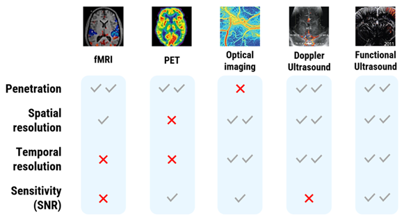
Image Credit: Iconeus
Functional ultrasound imaging (fUSI), like conventional Doppler ultrasound, is based on neurovascular coupling. When neurons are active, local blood vessels dilate, increasing cerebral blood volume (CBV).
Ultrafast Doppler sequences detect CBV changes with high sensitivity; however, unlike conventional Doppler, which scans tissue sequentially, fUSI images entire planes at thousands of frames per second (Mace et al., 2011, Nat Methods).
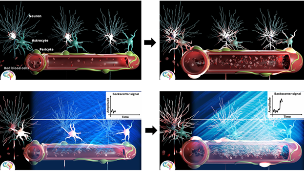
Image Credit: Iconeus
This strategy produces:
- High spatial resolution (100 µm) and temporal resolution (100–400 ms).
- Detection of CBV variations as small as 2 % from baseline.
- The signal-to-noise ratio is sufficient to map fine-scale vascular dynamics. Captures blood arteries with velocities as low as 1 mm/s (small arterioles).
These characteristics make fUSI ideally suitable for studying drug-induced effects across the entire brain in preclinical models.
Preclinical applications: Using fUS from model validation to therapeutic monitoring
Validating a disease model is a vital stage in preclinical research, but it is sometimes overlooked, despite its usefulness in evaluating therapy methods.
Functional ultrasound imaging (fUS) is an effective method for cross-validating models by observing neuronal and hemodynamic responses, functional connectivity changes, and vascular structural changes due to its sensitivity, spatiotemporal resolution, and whole-brain coverage.
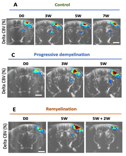
Image Credit: Iconeus
Beliard and colleagues (Imaging Neuroscience, 2025) provided a clear demonstration by employing fUS to define a cuprizone-induced model of multiple sclerosis.
Their tests replicated the demyelination-induced increase in hemodynamic responses in primary sensory cortices previously seen in MS patients using fMRI. Importantly, remyelination restored the baseline vascular signals.
The scientists further demonstrated that the CBV increase represented cortical vascular anomalies rather than demyelination alone, and that these alterations were missing in subcortical regions or in lysolecithin-induced localized lesions, indicating a cortex-specific mechanism associated with oligodendrocyte loss.
Beyond model validation, fUS is ideally adapted to characterizing the functional and vascular characteristics of neurological and neuropsychiatric illnesses, revealing how diseased processes modify brain activity and network dynamics, which is critical for developing targeted therapeutics.
Altered resting-state functional connectivity (FC) is a common trait across diverse contexts, and its cross-species conservation makes it an important readout in translational studies.
In a rat model of osteoarthritis, Rahal and colleagues used fUS to detect widespread reductions in connectivity across somatosensory and hippocampal networks.
Dynamic FC analysis revealed that arthritic animals spent much more time in states in which sensory cortical areas were disconnected from the rest of the somatosensory network.
When these connection metrics were associated with behavioral pain scores, the authors identified a set of areas that served as predictive biomarkers, enabling highly accurate differentiation between healthy and arthritic animals.
These findings emphasize fUS's potential to track network plasticity and identify functional characteristics relevant to therapeutic intervention.
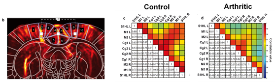
Image Credit: Iconeus
Once a model has been validated and any malfunctioning circuits have been discovered, fUS provides a reliable platform for screening and monitoring pharmacological effects.
Recent research has demonstrated that fUS can detect drug-specific "network fingerprints," distinguishing therapeutic actions from adverse effects, for example, in assessing opioid-induced changes in brain connectivity (Mariani et al., 2024).
Another key example comes from Rabut et al. (NeuroImage, 2020), who developed the concept of pharmaco-fUS to measure acute pharmacological effects in awake mice using whole-skull imaging.
They used machine-learning-derived pharmacological scores to accurately distinguish scopolamine therapy from baseline and found dose-dependent alteration of the hippocampo-cortical connection.
Importantly, these effects were independent of global perfusion alterations, demonstrating the specificity and sensitivity of fUS in pharmacological investigations.
These examples demonstrate how fUS offers a comprehensive preclinical workflow, from illness model development to circuit dysfunction analysis and therapy response monitoring using quantitative biomarkers based on CBV, activation mapping, and connectivity.
Translational potential and clinical outlook
Despite fUS imaging's well-established usefulness in preclinical neuroscience, several recent studies have demonstrated its translational utility in humans.
fUS has previously been used in intraoperative neurosurgery, newborn brain imaging, and epilepsy monitoring to create high-resolution vascular and activation maps in clinical settings.
These early applications demonstrate striking parallels with fMRI and electrophysiology, highlighting the potential to translate preclinical findings into patients.
Despite the clinical application still being in its early stages, these findings highlight the importance of fUS as a modality that connects preclinical models and human physiology, thereby increasing its usefulness for therapeutic development.
Iconeus Solutions for neuropharmacology research
High-quality neuropharmacology investigations require more than just sensitivity; they also require integrated instruments that enable exact acquisition, rigorous quantification of drug effects, and fully reproducible analysis. Iconeus offers a comprehensive methodology designed specifically for preclinical drug development and brain-targeted therapies.
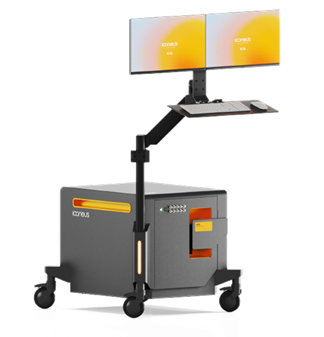
Image Credit: Iconeus
Hardware
- The Iconeus One system is a portable imaging station with ultrafast plane-wave technology and a fully integrated workstation. A height-adjustable keyboard and monitor provide comfortable operation, while a four-axis motorized scanning platform enables steady, repeatable imaging in head-fixed animals.
- The multi-array probe captures four planes concurrently, enabling whole-brain mice coverage in 2.4 seconds for quick assessment of drug-induced hemodynamic changes. It can be used transcranially or with craniotomy.
- The RCA probe's row-column technology enables rapid volumetric imaging of an 8 × 8 × 20 mm brain volume in 400 ms, making it ideal for capturing rapid cerebrovascular responses to drugs.
Software
- IcoScan is a user-friendly acquisition tool designed for fUSI drug investigations, providing stable baselines, dosing paradigms, and real-time monitoring of hemodynamic responses.
- IcoStudio is an advanced analysis environment that supports neuropharmacological procedures, including relative CBV measurement, activation maps, drug response dynamics, and functional connectivity analysis.
- IcoLab is a batch and group-level processing tool that promotes repeatability across cohorts and dosage settings. It incorporates filtering and artifact-rejection procedures and produces summary reports (in PowerPoint) as well as exportable datasets for further statistical analysis.
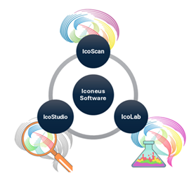
Image Credit: Iconeus
Together, these tools enable a smooth transition from acquisition to publication-ready results, transforming fUSI into a robust and accessible platform for researching drug-induced effects across the brain.
WANT TO LEARN MORE? Click here to watch the video
About Iconeus
Iconeus is a Paris-based company, founded by the inventors of functional ultrasound, who have invented an easy-to-use functional ultrasound system for imaging cerebral blood flow and microvasculature. Its unique combination of sensitivity, speed and high resolution has enabled it to lead the field in preclinical studies on awake animals, and it is now being proposed for applications in clinical research too.
Iconeus is introducing functional ultrasound neuro imaging: a breakthrough imaging modality for brain activity monitoring based on blood flow imaging with ultra-high sensitivity.
For preclinical research, the portability and high versatility of their technology enables the study of brain activity at unprecedented scales and in a large variety of subject states: awake, behaving, freely-moving, resting-state and asleep conditions.
Iconeus' technology can be easily combined with other complementary modalities such as EEG headstage or optogenetics which are inherently difficult to combine with FMRI. Iconeus also offers key applications such as functional connectivity assessment between brain structures (connectomics) or stroke 4D monitoring. Building on years of preclinical expertise and research, they're now investing and supporting exciting new clinical applications.
Sponsored Content Policy: News-Medical.net publishes articles and related content that may be derived from sources where we have existing commercial relationships, provided such content adds value to the core editorial ethos of News-Medical.net, which is to educate and inform site visitors interested in medical research, science, medical devices and treatments.