Diamond Light Source is one of the world’s most advanced scientific facilities. Located at the Harwell Science and Innovation Campus in Oxfordshire, it houses the UK’s national synchrotron facility alongside state-of-the-art cryo-electron microscopy technology.
Diamond operates as a not-for-profit organization, providing national infrastructure that supports leading research across the life sciences and materials sciences.
The synchrotron at Diamond functions much like a giant microscope and is used across a wide range of fields, including health, environmental science, engineering, and astrophysics.
Around ten thousand times more powerful than a traditional microscope, it accelerates electrons to near-light speed to generate exceptionally bright beams of light, known as beamlines.
These beams offer high brilliance, intensity, sharp focus, and tunability, enabling researchers to study samples in near-physiological conditions with remarkable detail.
World-class imaging under near physiological conditions
Diamond provides the scientific community with access to a broad range of equipment and laboratory facilities, including the Biological Imaging Center, which delivers advanced electron microscopy capabilities supported by specialist expertise.
Its biological cryo-imaging beamline, Beamline B24 full-field transmission microscope, was developed in response to growing demand for tomographic imaging of biological specimens under near-physiological conditions.
Beamline B24 brings together two instruments: the transmission cryo-soft X-ray tomography (cryoSXT) end station and the super-resolution fluorescence cryo-structured illumination microscope (cryoSIM).
The cryoSIM is an advanced cryogenic microscopy platform developed by Diamond in collaboration with research groups across Europe and Linkam Scientific Instruments. Used in tandem, these systems allow researchers to gain deeper insight into the structure and function of cells and tissues.
Dr. Harkiolaki summarises the team’s goals: “Current commercial fluorescence microscopes have, until now, been limited by low resolutions. For the first time, the cryoSIM allows correlation of super-resolution fluorescence imaging of cryopreserved specimens with their cellular ultrastructure. At Diamond, we offer the scientific community a critical mass of expertise in experiment design and data interpretation, together with access to world-class instrumentation to help solve a wide range of research questions. Our own research at the facility focuses on continuous innovation to address scientific challenges.”
Introducing the powerful cryoSIM
Imaging cells or tissues at different scales requires clear, unambiguous data correlation from the macroscopic level down to the microscopic.
One of the main challenges in high-resolution cellular imaging, however, is preserving native cell structures. This has led to the adoption of rapid cryo-preservation as the current gold standard for preparing biological samples for X-ray imaging.2
Existing cryogenic fluorescence localization methods have not been able to provide comparable resolution, limiting direct, like-for-like data comparison. In response, Diamond developed a bespoke microscopy solution that combines two technologies, enabling precise 3D data correlation for more detailed analysis.
A key part of this process is the plunge-freeze function of the cryoSIM, integrated directly into the microscopy stage. The cryogenic environment preserves the specimen (or even a dynamic reaction within it), maintaining native architecture and effectively capturing a snapshot of cellular activity at a defined moment in time.
The cryoSIM itself is a custom-built microscope based on a commercial cryogenic microscopy stage from Linkam Scientific Instruments. It enables 3D super-resolution fluorescence cryo-imaging and addresses many of the technical challenges associated with fluorescence imaging under cryogenic conditions, while doubling achievable resolution in all three dimensions.
Because the facility already had a Linkam stage for conventional cryo-microscopy imaging, the team chose to continue the collaboration, designing the custom cryoSIM according to the same principles.
This approach also allows for sample grid transfer and accurate data correlation between the two microscopy systems.
Together, these capabilities support the thoughtful design of fluorescence-dependent experiments and provide deeper insight into the inner workings of biological material.
Dr. Harkiolaki comments: “We have worked with Linkam for eight years, and the team’s highly collaborative approach brings considerable value to our work. Linkam is led by engineers, who know their product and understand the field inside out, and that makes a huge difference. It is not only that the business is heavily invested in microscopy – it is also that the company ethos is built around listening and responding to specific market needs with custom-designed products. Linkam has always been very responsive to our requirements – the team really took on our brief and has worked alongside us to help create exactly what we needed.”
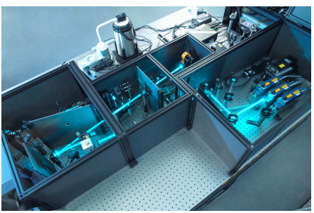
Figure 1. CMS196 stage setup incorporated in a cryoSIM at the Diamond Light Source Facility. Image Credit: Diamond Light Source
Cutting-edge imaging technology
The original aim of Beamline B24 was to generate 3D maps of cellular ultrastructure at a resolution of 25 nanometers. Although this does not match the resolution achieved by electron tomography, it offers a clear advantage in that a larger field of view captures the entire cell.
The next stage in its development focused on identifying the chemical localization of cells within these 3D ultrastructural maps. To achieve this, the cryoSIM microscope was developed using the structured illumination principle.
Structured illumination is built around the concept that the two microscopy systems enable seamless sample transfer for further, more detailed analysis without compromising data precision.
By using the same transmission electron microscopy (TEM) grid across both systems (first mapping the sample on one microscopy stage, then transferring it directly to the cryoSIM for 3D super-resolution fluorescence imaging), the resulting datasets can be overlaid accurately and directly onto the same sample.
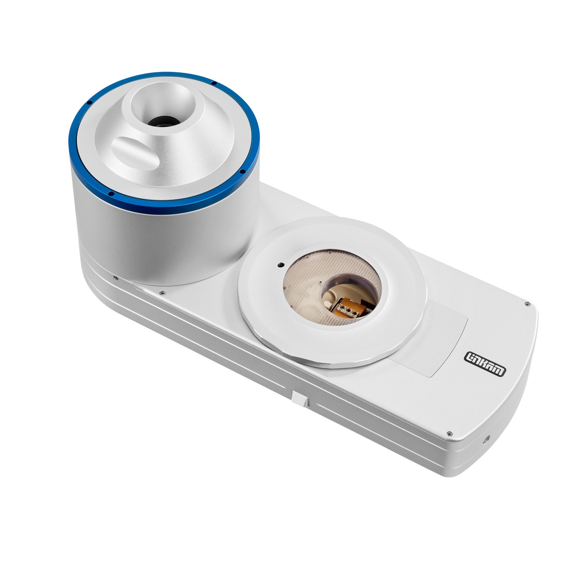
Figure 2. The Diamond Light CryoSIM allows 3D super-resolution fluorescence cryo-imaging, using a custom-built microscope based on the CMS196 stage from Linkam Scientific Instruments. Image Credit: Linkam Scientific Instruments
This integrated two-system approach yields a chemical signature as well as ultrastructure and content information, resembling a computerized tomography (CT) image of a cell that highlights key locations. This combination generates a 3D map of a cell and the location of any chemical entity within that map.
Dr. Harkiolaki explains: “Correlation usually has to be inferred from one sample to another, because the same sample could not, until now, be taken to different microscopes.
Now, by working with the same sample, I know for a fact that what I am seeing in the cryoSIM in fluorescence is the exact same area that I am seeing in the X-rays. This is a major step forward in cell sample imaging.”
Enabling in-depth imaging
The capability of this novel imaging platform has been demonstrated in a study of reovirus infection in human cells during the early stages of infection.3
The research combined cryo-structured illumination fluorescence imaging with soft X-ray absorption imaging on the same sample, enabling direct data correlation and opening up new opportunities for biological insight.
These high-resolution techniques enabled datasets to be overlaid in 3D on the same cell samples, allowing tracking of events as infection progressed through its early phases.
A subsequent study demonstrates that super-resolution imaging has changed fluorescence microscopy in fixed samples and emphasizes the effectiveness of several live cell imaging experiments.4
The findings demonstrate that improved resolution enhances the informational value of the correlative approach by enabling more precise localization of specific molecules such as proteins and nucleic acids within the context of ultrastructural morphology.
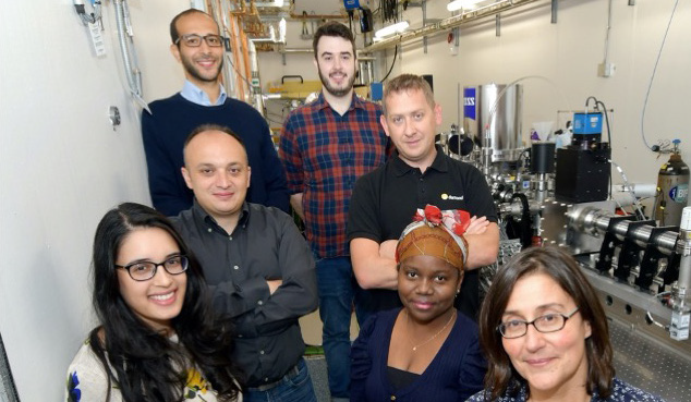
Figure 3. Dr. Harkiolaki (front right) and the team at Diamond Light Source, Oxfordshire. Image Credit: Linkam Scientific Instruments
Super-resolution imaging shows promise in new cancer therapies
The combined microscopy approach is also being applied in emerging cancer research.
One study investigating photoactivatable platinum (Pt) anticancer complexes for chemotherapy used synchrotron-based techniques to examine the in-cell behavior of these prodrugs.
For the first time, researchers were able to visualize changes in cellular morphology and platinum localisation following treatment, both with and without light irradiation.5
Photoactivated chemotherapy (PACT) works by selectively activating otherwise inert molecules, generating a localized antiproliferative effect. It is already used in the treatment of certain cancers, including skin, prostate, and bladder cancer.6
The study used cryo-SXT, nanofocused X-ray fluorescence (XRF), and X-ray absorption near-edge structure (XANES) to look into the anticancer mechanism of action of photoactivatable Pt (IV) PACT agents in their near-active form.
It was found that “The primary advantage of the method within the context of this study is that the setup at Beamline B24 has been constructed specifically to accommodate cryopreserved samples that can be used to collect further data in other microscopes … and therefore allow the unambiguous association of variable-contrast imaging data from the same sample.”7
Uncovering new biological insights
Dr. Harkiolaki concludes: “We are proud to deliver new innovations that support increasingly advanced research. Our own work is currently looking at various streams, including research into influenza, where we have seen excellent results on the egress pathway. We are also investigating cell-to-cell communication and responses to pathogen interactions.”
“Our success is driven by open collaboration, innovation, and the ability to engage in dialogue. We need people who can work together to support our goals, and Linkam as a partner fits that profile. Throughout our work, we aim to offer researchers world-class technology and expertise, which is demonstrated by our advanced imaging techniques unlike any other currently available.”
“Working with Linkam has definitely supported us in achieving our goals, and the team has been a key contributory factor in the success of the cryoSIM. We look forward to working on future projects together.”
Acknowledgments
This article is based on material originally authored by Dr. Maria Harkiolaki, Principal Beamline Scientist at Diamond Light Source.
References
- Collaboration in 2020 between Diamond Light Source and research groups and facilities across Europe: University of Oxford, UK, Heidelberg University Hospital, Germany, Université de Nantes, France, CryoCapCell, Paris, France and Micron, which is based at the Department of Biochemistry at the University of Oxford
- Koning, R. I., Koster, A. J., and Sharp, T. H. (2018). Advances in cryo-electron tomography for biology and medicine. Annals of Anatomy - Anatomischer Anzeiger, 217, 82–96. DOI: 10.1016/j.aanat.2018.02.004. https://www.sciencedirect.com/science/article/pii/S0940960218300219.
- Ilias Kounatidis, et al. (2020). 3D Correlative Cryo-Structured Illumination Fluorescence and Soft X-ray Microscopy Elucidates Reovirus Intracellular Release Pathway. 182(2), pp.515-530.e17. DOI: 10.1016/j.cell.2020.05.051. https://www.cell.com/cell/fulltext/S0092-8674(20)30684-X.
- Phillips, M.A., et al. (2020). CryoSIM: super-resolution 3D structured illumination cryogenic fluorescence microscopy for correlated ultrastructural imaging. Optica, 7(7), pp.802–802. DOI: 10.1364/optica.393203. https://opg.optica.org/optica/fulltext.cfm?uri=optica-7-7-802.
- Bolitho, E.M., et al. (2021). Single-Cell Chemistry of Photoactivatable Platinum Anticancer Complexes. Journal of the American Chemical Society, 143(48), pp.20224–20240. DOI: 10.1021/jacs.1c08630. https://pubs.acs.org/doi/10.1021/jacs.1c08630.
- Imberti, C., et al. (2019). New Designs for Phototherapeutic Transition Metal Complexes. Angewandte Chemie International Edition, 59(1), pp.61–73. DOI: 10.1002/anie.201905171. https://onlinelibrary.wiley.com/doi/10.1002/anie.201905171.
- Bolitho, E.M., et al. (2021). Single-Cell Chemistry of Photoactivatable Platinum Anticancer Complexes. Journal of the American Chemical Society, 143(48), pp.20224–20240. DOI: 10.1021/jacs.1c08630. https://pubs.acs.org/doi/10.1021/jacs.1c08630.
About Linkam Scientific Instruments
Linkam develops and manufactures a broad range of temperature and environmental control stages for both OEMs and end-users. From high to cryo temperatures as well as humidity, electrical connections, gas purging, vacuum, and pressure, for enhanced sample analysis. Linkam stages are used with light microscopes and a wide range of analytical techniques including Raman, FTIR, WAX/SAX, and many more to visualize and characterize the properties of materials.
Linkam has stages for research applications including Pharmaceutical, Chemical, Biological, Materials Science, Geology, Food Science and many more. The hot stages, environmental control add-ons, electronics, and software are all designed and built on-site thereby enabling us to quickly respond to suggestions made by current Linkam users, and so enabling us to continually develop and improve our designs as well as offering custom solutions.
Linkam stages are found in thousands of laboratories worldwide with the most successful microscope heating stage, the THMS600, selling over 6,000 units alone. Linkam is the market leader in temperature-controlled microscopy.
Sponsored Content Policy: News-Medical.net publishes articles and related content that may be derived from sources where we have existing commercial relationships, provided such content adds value to the core editorial ethos of News-Medical.net, which is to educate and inform site visitors interested in medical research, science, medical devices and treatments.