Multiplex immunohistochemistry (mIHC) involves multiple staining and imaging steps, meaning problems can arise at different stages of the workflow. Troubleshooting is therefore most effective when it begins with the observed symptom, then considers its likely cause and the appropriate corrective action.
In particular, incomplete HRP quenching and suboptimal antibody concentrations are common sources of problems and should be among the first factors considered when more than one symptom is present.
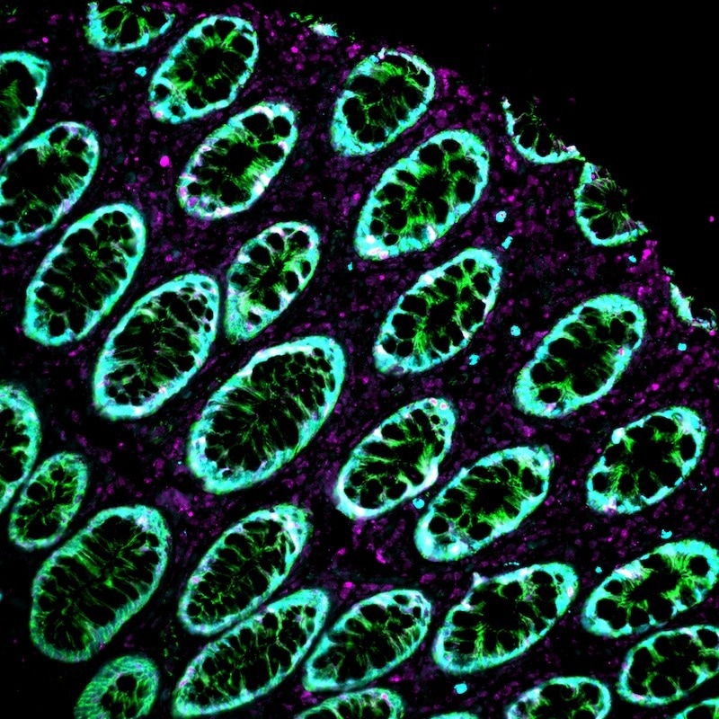
AtlasPlex 3-plex, human rectum. Image Credit: Atlas Antibodies
Signal and detection
When there is no signal on a target, a likely cause is insufficient antigen retrieval for the target, or a primary antibody concentration that is too low. In such a situation, the antibody should be titrated upward in two-fold steps. An alternative antigen retrieval buffer can be tested, for example, switching from citrate pH 6 to EDTA pH 9. Before the antibody is multiplexed, the user should confirm that it works in a standard single-plex.
Targets with low abundance present a unique challenge. Often, this is a result of the target being placed in a late cycle where background baseline has begun to accumulate. Moving the target to cycle 1 or 2 could help to address this problem. As in the previous issue, a single-plex control should be run before diagnosing a panel design issue.
When moving between a pilot and a full panel, signal can be lost. Sometimes, this is due to epitope degradation or tissue morphology following HRP quenching in a previous cycle. Users should check quenching time and concentration; reduce the duration of quenching; and confirm tissue integrity on an H&E from the same block.
At the other end of the spectrum, negative control tissue can have a strong signal. Two key causes of this are non-specific antibody binding and endogenous binding/peroxidase activity. To overcome this, users should increase blocking time or concentration. If using biotin-rich tissues, such as liver, kidney, and brain, avidin-biotin blocking can be added. Finally, the secondary-only control must be clean.
Background and noise
Inadequate blocking or non-specific TSA deposition can cause a high background across the entire section of tissue. Three methods can be used to fix this: extending the blocking step; reducing TSA reagent concentration; and ensuring the HRP is fully quenched before the next cycle. This final method is perhaps the most helpful, since incomplete quenching is the most common cause of background accumulation.
Sometimes, the background can increase with each cycle, due to incomplete HRP quenching between cycles. In response, quenching incubation should be extended, the freshness of the quenching reagent should be verified, and a no-primary control should be run for each cycle–this will help identify which cycle is the source.
Another key issue is when bright punctate spots appear across the tissue, which points to aggregated antibody or reagent precipitation. Antibody solutions can be centrifuged before use (10,000 g for five minutes) to address this, and the TSA working solution should be filtered. Freeze-thaw cycles of concentrated antibody stocks are to be avoided.
Specificity
If the wrong antibody clone is chosen for IHC or poor fixation causes protein leakage, the nuclear marker may show cytoplasmic signal. The optimal time for fixation is 24–48 hours in NBF. If this time is adhered to but issues ensue, the user should confirm the antibody has published IHC validation data and run the sample on tonsil or another appropriate positive control tissue.
To fix the issue of nuclear markers bleeding into one another, likely caused by markers being placed in consecutive cycles with spectrally close fluorophores, they should be separated with a non-nuclear marker cycle. To ensure nuclear channels are spectrally distinct, fluorophores can be reassigned.
Cases of unexpected co-expression on the same cell may reflect true biology or cross-reactivity between antibodies. In this case, antibodies must be run as single-plex on consecutive sections; if co-expression occurs only in the multiplex panel, antibody cross-reactivity and epitope competition should be investigated.
Imaging
A key imaging concern is channel bleed-through on imaging. Since this is likely caused by non-fully corrected fluorophore spectral overlap, single-fluorophore reference images should be acquired for each channel and imaging software should be spectrally unmixed. If bleed-through persists, spectral separation can be increased by reassigning fluorophores.
If antibodies are unevenly distributed during incubation or tissue drying, the slide’s signal intensity can also become uneven. To prevent this, sections must be fully covered during incubation: a humidity chamber can help. In early cycles, it is especially important to ensure that the tissue’s edges do not dry up.
A final symptom is tissue detachment, which is often caused by insufficient section adhesion to the slide or wash steps that are overly aggressive. SuperFrost Plus or poly-L-lysine-coated slides can help, as can baking sections at 60 °C for one hour before starting and reducing the wash agitation speed.
About Atlas Antibodies AB
Atlas Antibodies, founded on the Human Protein Atlas project, provides a comprehensive range of highly validated antibodies targeting over 75% of the human proteome. Core products include Triple A Polyclonals™ advanced rabbit polyclonal antibodies, and PrecisA Monoclonals™ precise mouse monoclonals designed for superior specificity.
Atlas Antibodies also offers PrEST Antigens™, recombinant protein fragments used for control experiments, and MolBoolean™, an innovative technology for in situ protein-protein interaction studies. Manufactured in Sweden and distributed globally, Atlas Antibodies’ products support diverse research applications with exceptional accuracy and reliability.
Sponsored Content Policy: News-Medical.net publishes articles and related content that may be derived from sources where we have existing commercial relationships, provided such content adds value to the core editorial ethos of News-Medical.net, which is to educate and inform site visitors interested in medical research, science, medical devices, and treatments.