Automated high content image acquisition and analysis for drug discovery and cell biology
The CELENA® X High Content Imaging System is an integrated imaging system designed for rapid, high content image acquisition and analysis. Customizable imaging protocols, image-based and laser autofocusing modules, and a motorized XYZ stage simplify well plate imaging and slide scanning. The integrated CELENA® X Cell Analyzer software processes images and data for quantitative analysis. Analysis pipelines can be put together and reused to identify cellular or subcellular objects, process images for optimal data collection, and make various measurements.
The CELENA® X is as flexible as it is powerful, with interchangeable objectives and filter cubes to accommodate a wide range of fixed and live cell imaging applications.
Simple High Content Imaging for Quantitative Image-Based Analysis
Fully Automated Plate and Slide Imaging
- Automated vessel handling and scanning
- Motorized XYZ stage, filter cube stage, and objective turret
Laser Autofocus
- Rapid and reproducible focusing
- Minimized phototoxicity and photobleaching
Live Cell Assay Support
Onstage incubation system for a variety of experiments in physiological and non-physiological conditions
Four Imaging Modes
Fluorescence imaging in four channels, brightfield, color brightfield, and phase contrast imaging
Powerful, Easy-To-Use User Interface
- Simple setup of imaging protocols
- Seamless integration of imaging and data analysis processes
Customizable High Content Analysis
- Create and customize image analysis projects
- Quantitatively analyze multiple image-based phenotypes
Create Your Own Imaging and Analysis Workflows with the CELENA® X
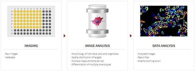
Abundant Applications
The CELENA® X Cell Analyzer software is incredibly powerful and its ability to batch process makes it ideal for high content analysis.
The powerful and flexible software allows for the creation and customization of workflows that can be used for the simplest fixed cell assays to more complicated, time-lapse live cell assays.
- Apoptosis
- Autophagy
- Cell cycle
- Cell counting
- Cell growth/proliferation
- Cell migration
- Cell morphology
- Cell viability/toxicity
- Co-localization
- Confluency
- Cytotoxicity
- Histology
- Immunofluorescence
- NF-κB translocation
- Organoid/spheroid morphology
- Transfection efficiency