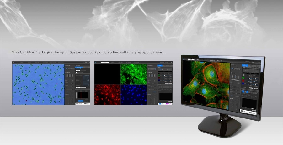
The CELENA® S Digital Imaging System is a comprehensive solution for capturing publication-quality fluorescence, brightfield and Phase contrast images. Sophisticated software accommodates a wide range of imaging applications such as image capture and analysis, live cell imaging, and even automated cell counting. Capture amazing detail with a few clicks of your mouse.
Imaging to data analysis with one device, in one sitting
The CELENA® S is a small and powerful digital imaging system that simplifies imaging and data analysis. Integrating advanced precision optics, a highly sensitive scientific grade CMOS camera, and a computer with user-friendly software, the CELENA® S allows researchers to capture vivid, publication quality images with ease.
Interchangeable objectives and filter cubes accommodate a wide range of imaging needs. Researchers can use the CELENA® S for multiple applications, such as capturing and analyzing multicolor fluorescence images, live cell imaging, and automated cell counting.
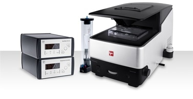
Onstage incubation system
Composed of an environmental chamber, temperature controller, and a gas mixer, the new onstage incubation system supports various live cell imaging applications. Researchers can control the temperature, humidity, and gas content with precision. Live cells can be monitored with the time lapse function or the growth monitor on the CELENA® S.
Features
All-in-one system
Combining a microscope, camera, light source, and a computer with software in one sleek package, the CELENA® S takes you from imaging to data analysis in one sitting.
Multicolor fluorescence and brightfield imaging
Long-lasting LEDs and hard-coated optical filters ensure robust fluorescence imaging. Adjustable LEDs allow precise control over the gain and intensity of transmitted light.
Onboard data analysis
Analyze your images immediately upon capture. Save measurement data to a USB drive.
Live cell monitoring
Monitor live cells with the time lapse function or the growth monitor. Attach the onstage incubator to control the temperature, humidity, and CO₂/O₂ levels.
Automated cell count and viability analysis
Check cell counts and viability with the onboard cell counter
Z-stack imaging
Capture multiple images along the Z-axis with the Z-stack function.
| CELENA® S Digital Imaging System Specifications |
| Imaging methods |
Epifluorescence and transmitted light (brightfield and phase contrast) |
| Illumination |
LED filter cubes with adjustable intensity (>50,000 hr life per filter cube) |
| Fluorescence channels |
3 fluorescence channels and 1 transmitted light channel. |
| Objective turret |
5 positions |
| Objectives |
High quality long working distance (LWD) and coverslip-corrected; 1.25X-100X |
| Condenser |
47 mm LWD condenser; 3-positions with brightfield and phase contrast annuli |
| Computer |
Built-in dual core CPU, 128 GB SSD internal storage |
| Stage |
Mechanical X/Y stage, motorized Z stage; accommodates an onstage incubator |
| LCD display |
Full HD color LCD monitor, 1920 x 1080 pixels (not included) |
| Cameras |
1.3 MP monochrome CMOS with 1280 x 1024 pixels |
| Images |
8 or 16-bit TIFF, JPG, BMP, or PNG |
| Dimensions (L x W x H) |
44 cm x 30 cm x 27 cm (17.3″ x 11.6″ x 10.6″) |
| Weight |
20 kg (44 lb |