From SemplorReviewed by Olivia Frost
This article is based on a poster originally authored by Dr. Ivan Lobato, Eric Toering, Dr. Serge Gavras, and Prof. Dr. Karel van der Mast from Semplor.
EDS spectra feature distinct X-ray peaks overlaid on a Bremsstrahlung continuum. The peaks reveal elemental makeup, while the continuum provides complementary data tied to the local mean-Z and sample geometry. With low counts, signal-dependent Poisson noise hides faint peaks and hinders quantitative spectral reconstruction.
Detector physics introduces additional ambiguities regarding structure. Si-escape peaks appear 1.74 keV beneath energetic parent lines, while pulse pile-up at high count rates produces sum peaks at E1 +E2. Sequential denoising and background subtraction fail to maintain the distinct peak and continuum components or clearly resolve these detector stages.
Objective: Simultaneously denoise EDS spectra, separate characteristic peaks from Bremsstrahlung, and correct for Si escape and pile-up within a single physics-informed inverse problem.
Method: Physics-structured targets and one coupled network
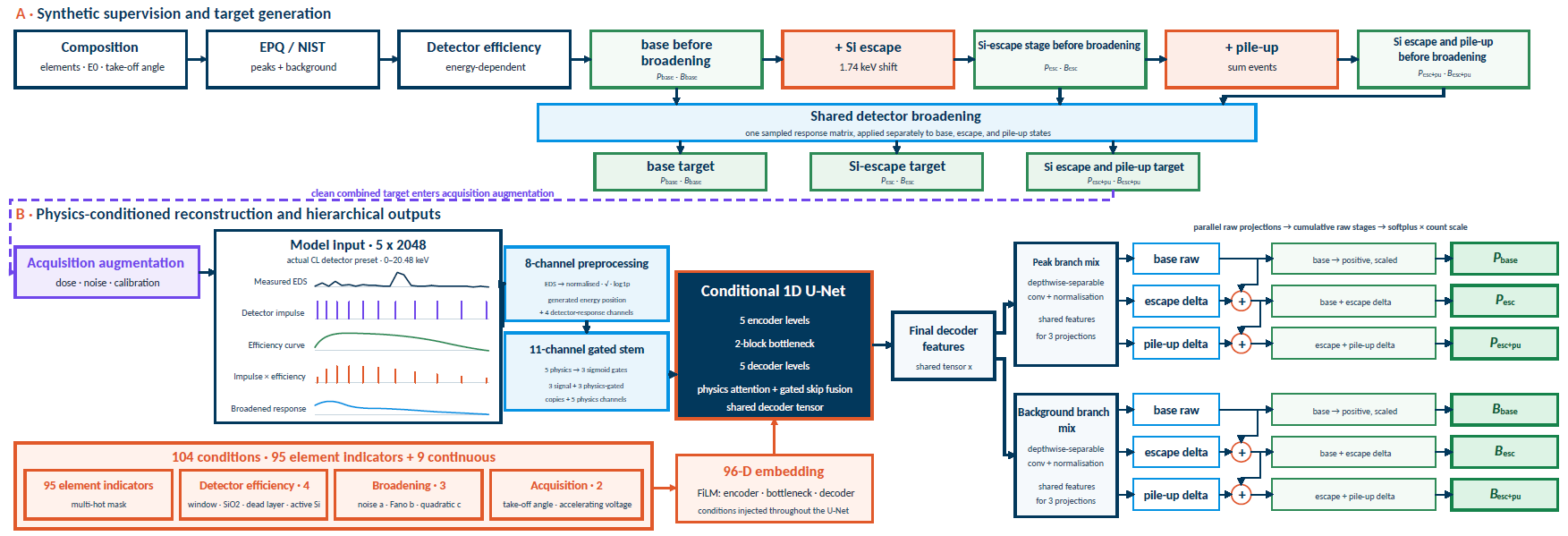
Image Credit: Semplor
Stage notation: P and B signify the peak and Bremsstrahlung components, respectively. Pbase/Bbase: Si escape and pile-up eliminated; Pesc/Besc: Si escape retained and pile-up removed; Pesc+pu/Besc+pu: both artifacts retained before stochastic acquisition augmentation.
Results 1: Fe2O3 spectra validate low-dose reconstruction and detector-stage separation
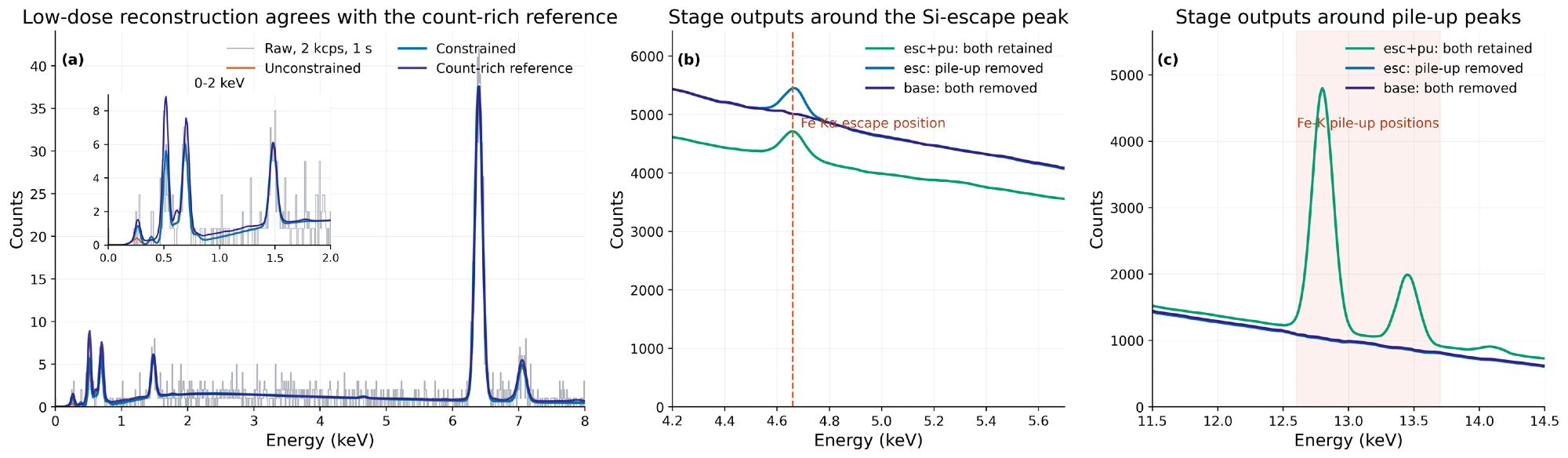
Image Credit: Semplor
At 20 kV, the constrained 2 kcps, 1 s reconstruction agrees with a count-rich reference. The stage-resolved outputs identify the Fe Kα Si-escape position and the Fe K pile-up region.
Results 2: Signal separation across the thermite spectral image
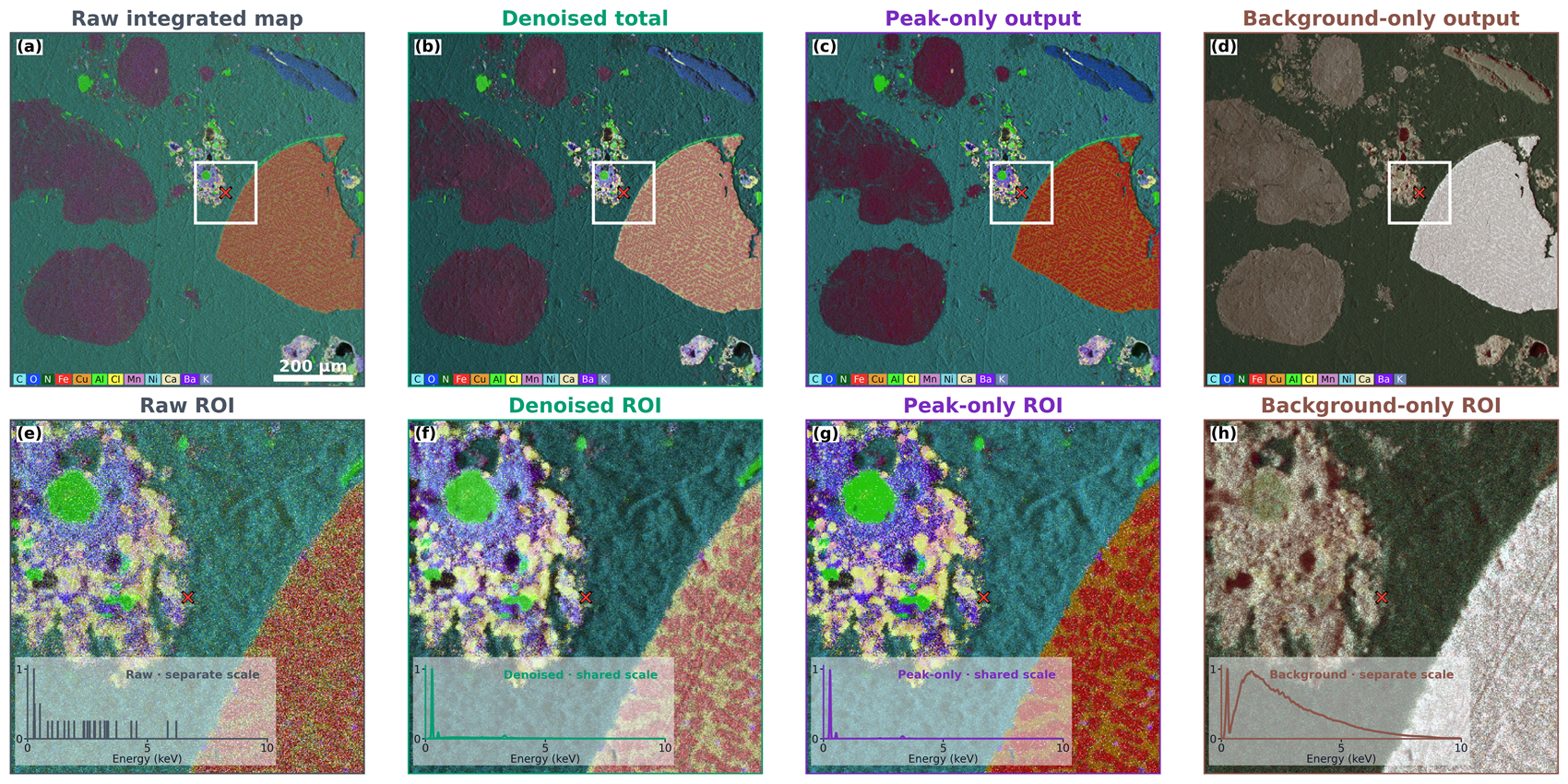
Image Credit: Semplor
Each column connects one full-field map with its designated ROI and central-pixel spectrum. Denoised and peak-only insets use the denoised-total scale; raw counts and background are normalized independently so their spectral shapes stay visible.
Conclusions
- Physics-structured targets, structured loss stages, and a shared conditional network distinguish low-dose denoising from compensation of detector artifacts; this approach is applicable across multiple acquisition conditions.
- Six coupled outputs from two hierarchical branches recover matched peak/background pairs at three cumulative physical stages: both Si escape and pile-up retained, Si escape retained with pile-up removed, and both artefacts removed.
- The experiments with Fe2O3 cover varying count ranges: conditioning enhances low-dose reconstruction, constrained and unconstrained solutions align as counts rise, while variations between stages highlight the Si escape and pile-up structure at elevated count rates.
- The thermite spectral imagery indicates that denoising reduces speckle, peak-only reconstruction enhances compositional differentiation, and the isolated background maintains complementary mean-Z and structural contrast. The model accommodates constrained, semi-constrained, and unconditional inference.
References and further reading
- Pouchou, J.-L. and Pichoir, F. (1991). Quantitative Analysis of Homogeneous or Stratified Microvolumes Applying the Model “PAP.” Electron Probe Quantitation, pp.31–75. DOI:10.1007/978-1-4899-2617-3_4. https://link.springer.com/chapter/10.1007/978-1-4899-2617-3_4.
- Ritchie, N.W.M. (2009). Spectrum Simulation in DTSA-II. Microscopy and Microanalysis, 15(5), pp.454–468. DOI:10.1017/s1431927609990407. https://academic.oup.com/mam/article-abstract/15/5/454/6919550.
- Schoonjans, T., et al. (2011). The xraylib library for X-ray–matter interactions. Recent developments. Spectrochimica Acta Part B: Atomic Spectroscopy, 66(11), pp.776–784. DOI:10.1016/j.sab.2011.09.011. https://www.sciencedirect.com/science/article/abs/pii/S0584854711001984.
- Ronneberger, O., Fischer, P. and Brox, T. (2015). U-Net: Convolutional Networks for Biomedical Image Segmentation. Lecture Notes in Computer Science, 9351, pp.234–241. DOI:10.1007/978-3-319-24574-4_28. https://link.springer.com/chapter/10.1007/978-3-319-24574-4_28.
- Semplor BV. NANOS tabletop SEM product information (2026).
About Semplor
Semplor is an innovative high-tech company based in Eindhoven, The Netherlands - the birthplace of modern electron microscopy. Eindhoven has long been a hub of innovation, with its roots in Philips Electron Optics during the mid-20th century.
Semplor was founded in 2021 by seasoned electron microscopy experts. The company’s flagship product, the NANOS, was developed by a team of passionate innovators with a deep understanding of EM technology, precision engineering, and a strong international network. Together, the team brings over 200 years of combined experience from leading companies such as Philips, FEI, Phenom, and Thermo Fisher Scientific.
Our mission is simple yet ambitious: to make Scanning Electron Microscopy (SEM) accessible for anyone, everywhere. Driven by passion and decades of experience, Semplor has created a new generation of tabletop SEM - compact, robust, and remarkably capable. During the development of the NANOS, we combined the latest insights in microscopy with smart, efficient engineering. The result is a high-performance, user-friendly instrument that delivers comprehensive imaging at an affordable cost. Engineered with dedication and expertise, the NANOS represents the next generation of tabletop SEMs - a perfect balance of quality, accessibility, and innovation.
Sponsored Content Policy: News-Medical.net publishes articles and related content that may be derived from sources where we have existing commercial relationships, provided such content adds value to the core editorial ethos of News-Medical.net, which is to educate and inform site visitors interested in medical research, science, medical devices and treatments.
Last Updated: Sep 30, 2026