An interview with Scott Olenych, from ZEISS, to discuss the challenges faced in neuroscience research microscopy techniques and to discuss their latest microscope that could help overcome some of these challenges.
Why did you exhibit at Neuroscience in 2019?
The annual Neuroscience meeting is a great place for Carl Zeiss Microscopy (ZEISS) to connect with research scientists to learn what research problems they are investigating and to understand how our microscopes can help them.
The Neuroscience meeting is one of the largest life science meetings in the USA and we look forward to attending each year. ZEISS has been attending Neuroscience for more than 20 years.
Are there any products you were promoting at the show?
This year at the Neuroscience meeting, we were pleased to exhibit the LSM 980 confocal microscope with Airyscan 2. This Airyscan 2 on an upright LSM configuration is a key part of a partnership that ZEISS has been developing for several years with Inscopix to enable researchers to use GRIN lens technology to image areas mouse brain that is unreachable with traditional in vivo microscopy.
The combination of Airyscan imaging through a GRIN lens can also be correlated with freely behaving imaging to provide additional context to the data.
ZEISS also demonstrated the Elyra 7 with Lattice SIM technology this year at Neuroscience. Lattice SIM is a Superresolution SIM imaging technique that provides optical sectioning with a doubling of diffraction-limited resolution (120 nm in xy and 300nm in z) while achieving up to 255 image frames per second. Lattice SIM has the speed and light efficiency to answer demanding high speed live Superresolution questions.

Image Credit: Zeiss.com
What are the challenges facing microscopy in Neuroscience research, treatment and development?
Major research questions in Neuroscience revolve around understanding brain structure and how structure relates to brain function. Microscopy helps to answer questions in both these areas by helping to visualize structural details of brain and cellular organization down to nanometer resolution over large areas of tissue and volume.
Imaging living brain and brain tissue activity offers new insights and can be also correlated with structural information. The challenges are to produce the resolution and detail needed to understand the interconnected areas of the brain while also imaging deeply with speed to capture fast events while also not damaging living cells and tissues.

Image Credit: Alexandros A Lavdas
How does Zeiss’ technologies help to provide high-contrast 3D and spectral imaging in neuroscience?
ZEISS is a leader in 3D imaging with a variety of light microscopy techniques such as laser scanning confocal microscopes, super-resolution techniques such as Lattice SIM, single-molecule localization microscopy (SMLM) and Airyscan.
These Light microscopy techniques can easily be used to collect multispectral imaging information. In electron microscopy (EM), ZEISS offers techniques that image sections of tissue milled with FIB-SEM, cut with a microtome inside the microscope or imaged as serial sections of tissue with high throughput. Imaging of serial sections with EM produces 3D data with resolutions in the sub 10 nm resolution range.
What new insights do Zeiss’ microscopes provide into a sample’s biology?
ZEISS microscopes provide highly detailed structural information as well as functional information. Images from a variety of microscopes, for example, from EM and light microscopes can be spatially correlated. Correlative microscopy has traditionally been very challenging, but new tools in ZEISS ZEN software are making this difficult task easier than ever before.
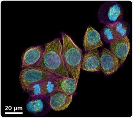
Image Credit: Zeiss.com
How does Zeiss offer the ability to image whole brain sections?
For cleared brain sections and whole small animal brains, ZEISS Lightsheet Z.1 is an excellent tool to choose from. The image acquisition speed of the Lightsheet Z.1 allows very fast imaging over large volumes of samples.
Lightsheet Z.1 offers multiple magnifications and images resolutions for whole-brain sections. Fixed and labeled whole brain sections could be imaged either with ZEISS Axio Scan.Z1 if thinly sliced and mounted on a slide or imaged with ZEISS LSM 900 or ZEISS LSM 980 if the section is thick and 3D information is needed.
How do you think the use of your technology will advance future scientific research and development? Can you provide any specific examples?
Thousands of ZEISS microscopes are used every day to aid in scientific discovery.ZEISS is continually advancing what is possible with microscopy to help scientists realize their goals.
What makes Zeiss and its technologies unique?
ZEISS was present at the very beginnings of research microscopy and was an early partner to scientists in helping to develop and commercialize innovative research microscopy techniques. ZEISS has continued this tradition of partnership for more than 150 years and has invested in and developed a portfolio of light microscopy, electron, ion and x-ray all focused on answering the difficult scientific questions.
About Scott Olenych.
Scott Olenych received his Ph.D. in Molecular Biology from Florida State University. His interest in fluorescent proteins and microscopy led him to the National High Magnetic Field Laboratory and later to Carl Zeiss Microscopy.
He has held a variety of positions at Zeiss and currently is a Product Marketing Manager for Carl Zeiss Microscopy, LLC in the United States.
 New Multiplex mode for ZEISS Airyscan 2 delivers images with superresolution in less time