In this interview, Sheela Muley, Product Manager at Molecular Devices, and Dr Sushmita Sudarshan, Application Scientist in Assay Development at Molecular Devices, talk about innovative approaches to microbiome research, with a focus on automation in anaerobic workflows. They discuss how next‑generation tools are helping to unlock the uncultured majority of the microbiome and streamline microbial screening for novel therapeutics.
Can you please introduce yourselves and your roles at Molecular Devices?
Muley: I’m Sheela Muley, Product Manager at Biopharma at Molecular Devices. In my role, I oversee the development and commercialization of microbial and mammalian clone‑screening platforms.
Dr Sudarshan: I’m Sushmita Sudarshan, Application Scientist in Assay Development at Molecular Devices. I work on translating microbial workflows into automated solutions, especially focusing on high‑throughput and anaerobic applications.
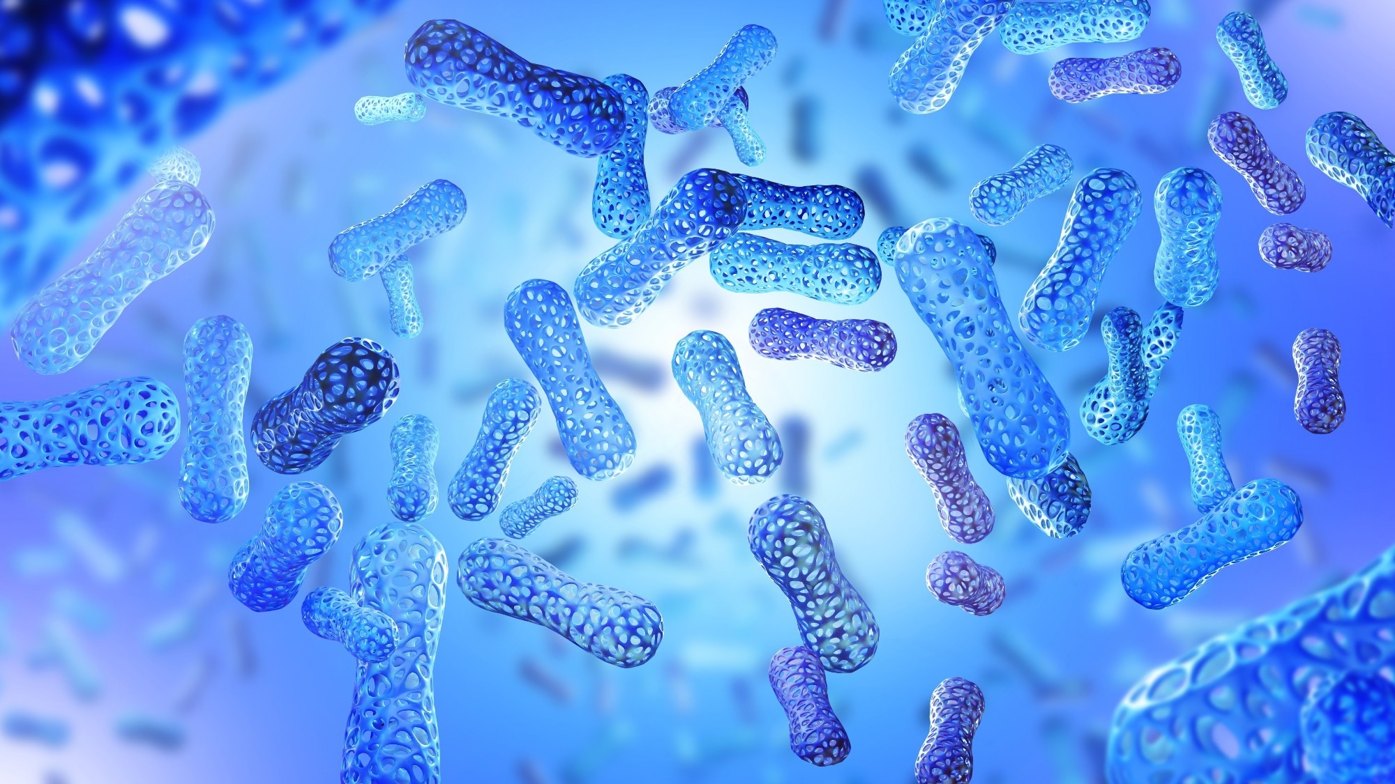
 Image Credit: FOTOGRIN/Shutterstock.com
Why is the microbiome such an important research frontier right now?
Muley: The microbiome is a key influencer of our immune system, metabolism, and even our mood and neurological health. We have trillions of microbes in our body, and they encode far more genes than our human genome. So, understanding that community and converting it into therapeutics has tremendous potential.
We also know that only about a third of gut microbial species are culturable today, meaning 60‑70% remain uncultured. That gap drives the need for new technologies and workflows.
We only know about 30 to 40 percent of our microbiome… so how do we get to that 100 % of this uncultured and unexplorable diversity?
Dr Muley, Product Manager at Biopharma at Molecular Devices
So, the microbiome is not just descriptive; it’s actionable if we develop the right tools.
What are the major challenges in anaerobic microbiome workflows?
Muley: A big challenge is culturing strict anaerobes. The gut and many environmental microbiomes are oxygen‑free or low‑oxygen habitats, and manual workflows inside anaerobic chambers are slow, error‑prone, and lack standardization and throughput. Preservation of sample integrity, sterility, and traceability is critical.
The functional gap here is going from genotype (NGS) to phenotype and interaction, which requires culturing, isolating, picking colonies, and so on.
How does the QPix FLEX‑system help address those challenges?
Muley: We developed our QPix FLEX microbial screening platform to automate plating, streaking, colony picking, hit‑picking, and liquid handling in a compact form factor compatible with anaerobic (hypoxic) chambers.
It includes features like a high‑resolution colour imaging camera for morphology and pigment detection, barcode tracking to reduce human error, sterilization modes (UV, ultrasonic baths, optional HEPA), the ability to use disposable tips, and a deck layout designed for flexibility.
We were able to pick three times more colonies within a week … saving one to three days of picking.
Dr Muley, Product Manager at Biopharma at Molecular Devices
By integrating multiple workflow steps in one instrument, we reduce instrument footprint, reduce sample exposure to oxygen, standardize protocols, and improve throughput.
Dr. Sudarshan, could you walk us through some of the platform's core automated processes and explain how they translate into time savings or consistency?
Dr Sudarshan: Absolutely. The four core processes we emphasize are:
- Colony picking (colour and morphology-based)
- Plating and streaking (onto agar trays, Omni‑Trays, Petri dishes)
- Liquid handling (via four‑channel expandable pipetting head)
- Hit‑picking / cherry‑picking (mapping defined colonies into master plates)
For example, our colour imaging enables grouping of colonies based on RGB intensity, helping differentiate pigmented or reporter strains (e.g., blue vs white on X‑gal media, or coliform differentiation on ECC agar)
These automated steps reduce manual steps, reduce errors, and ensure consistent volume dispensing (validated via absorbance). They also enable the selection of smaller colonies earlier, avoiding contaminants' overgrowth. That translates into days of time saved and increased colony yield and reproducibility.
How does the system maintain anaerobic‑friendly workflows and preserve sample integrity?
Dr Sudarshan: The system is purpose‑built for anaerobic integration:
- A compact footprint so it fits inside standard anaerobic or hypoxic chambers
- It avoids heat or compressor‑based sterilization, so picking pins can remain air‑dried (or use disposable tips) without introducing oxygen or heat shock to strains
- The system was tested inside a hypoxic chamber with 5% hydrogen for 1.8 years for stability
- Automation reduces the number of times plates are moved in/out of the chamber, reducing exposure to ambient air, contamination risk, and stress.
These design choices ensure we’re automating microbial workflows while preserving the viability of oxygen‑sensitive microbes and enhancing reproducibility.
The colour‑camera and colony morphology classification sounds powerful. Could you explain how that works and why it’s important?
Dr Sudarshan: The system uses a 20‑megapixel CMOS colour camera combined with intelligent software to generate RGB histograms for each colony image. Colonies can be classified based on colour intensity (red, green, blue channels) and morphology parameters (compactness, aspect ratio, diameter).
For example, a blue colony (LacZ positive) vs a white colony (LacZ negative) can be separated by their RGB profiles. The operator interface allows for the selection of a reference colony and the grouping of similar colonies automatically. The software includes thresholding algorithms (e.g., Fensalke local threshold) for high‑fidelity detection even under low contrast.
This means you’re not just picking by size or location, you can pick based on phenotype (pigment, expression marker), which is critical for microbiome workflows or engineered strains.
What genomics methods do you recommend for the isolates obtained through these high‑throughput workflows?
Muley: For microbial identification and genomic anal,ysis we typically use 16S rRNA sequencing for species‑level identification of anaerobic isolates. For functional or protein‑fingerprint data, MALDI‑TOF is useful (e.g., for strain ID post‑isolation). If you want both genotype and function, you could combine 16S (or full‑genome sequencing) with MALDI‑TOF or phenotypic assays. The key is linking genotype to phenotype in a systematic, reproducible way.
Looking ahead: How does automation in microbiome research reshape the future?
Dr Sudarshan: Automation enables labs of all sizes, not just large pharma, to participate meaningfully in microbiome culturomics.
By replacing manual, error‑prone workflows with automated, traceable ones, we make it possible to tap into the uncultured majority, accelerate probiotic discovery, and translate the microbiome into next‑generation therapeutics.
Muley: Indeed, the next breakthrough in a condition like Alzheimer’s, depression, or cardiovascular disease may come from the microbiome. But it requires robust workflows and automation to scale discovery. We hope to see automation become infrastructure rather than a luxury, facilitating faster, reproducible science across industry and academia.
Finally, what advice would you offer researchers embarking on anaerobic microbiome screening?
Muley: Start by thinking about the full workflow: sample collection (and maintaining anaerobic environment), plating, isolation, colony picking, tracking, and analysis. If your instrument or workflow only solves one step, you risk bottlenecks elsewhere.
Dr Sudarshan: Prioritize sterility, traceability, and automation early. Use barcoding, imaging, and integrated tracking so you’re not just picking colonies, you’re building reproducible datasets. And don’t wait to pick large colonies, automated imaging enables earlier picks, saving time and reducing contamination risk.
About the Interviewees
Sheela Muley
Sheela Muley is Product Manager, Biopharma at Molecular Devices, where she leads the microbial and mammalian clone‑screening portfolio. With over 20 years of experience in life‑science instrumentation, high‑throughput screening, and assay commercialization, Sheela Muley has overseen product initiatives spanning analytical platforms and cell‑based screening workflows.
She holds a doctorate in a relevant life‑science discipline (specific institution not public). Her expertise encompasses automation, instrumentation validation, and translating evolving technologies into market‑ready scientific tools. She regularly presents on emerging trends such as microbiome culturomics and anaerobic automation workflows.
Dr Sushmita Sudarshan
Dr Sushmita Sudarshan is Application Scientist in Assay Development at Molecular Devices, specialising in microbial research, assay automation, and lab instrumentation. She earned her PhD in Molecular Biology from the University of Texas at Dallas.
With over a decade of experience spanning academic and industry labs, she has developed and validated automated microbial workflows, including integration of high‑content imaging, colony picking, and traceable analytics. Her work supports bacterial, anaerobic, and synthetic biology applications across research and therapeutic development.
About Molecular Devices UK Ltd
Molecular Devices is one of the world’s leading providers of high-performance bioanalytical measurement systems, software, and consumables for life science research, pharmaceutical, and biotherapeutic development. Included within a broad product portfolio are platforms for high-throughput screening, genomic and cellular analysis, colony selection, and microplate detection. These leading-edge products enable scientists to improve productivity and effectiveness, ultimately accelerating research and the discovery of new therapeutics. Molecular Devices is committed to the continual development of innovative solutions for life science applications. The company is headquartered in Silicon Valley, California, with offices around the globe. For more information, please visit www.moleculardevices.com.