The stRAMos Photothermal Stimulated Raman Microscope employs the PhotoThermalSRS™ (PT-SRS) method to provide the utmost sensitivity in measurements and spatial resolution for chemical imaging within one system, featuring rapid imaging speeds and enhanced microscopy characterization capabilities.
- High-speed chemical imaging for live cells and molecular dynamics
- Highest sensitivity measurements: 10× improvement over SRS
- ≤300 nm Raman chemical spatial resolution
About stRAMos
The stRAMos microscope uniquely integrates a variety of optional multimodal features into one cohesive system. It facilitates the integration of PT-SRS with optical photothermal infrared spectroscopy (O-PTIR), widefield epi-fluorescence imaging, and spontaneous Raman spectroscopy, thereby enabling comprehensive chemical characterization of the sample under analysis.
stRAMos, featuring its multimodal functionalities, offers the most extensive characterization in chemical imaging and spectroscopy for various applications in the life sciences. This includes imaging of both live and dried cells, as well as the biomolecular dynamics of proteins, lipids, and other biomolecules.
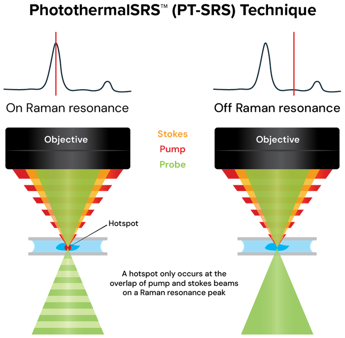
Image Credit: Photothermal Spectroscopy Corp
stRAMos key capabilities
The subsequent videos demonstrate the stRAMos system applied to yeast cells cultivated on sugar, resulting in enhanced lipid production. PT-SRS allows for rapid imaging of lipids and proteins within the cells.
Hyperspectral chemical imaging in live cells
PT-SRS enables comprehensive hyperspectral imaging across both the C–H and fingerprint regions, capturing abundant chemical differences without the need for labels. By obtaining spectra from each individual pixel, scientists are able to identify subtle yet significant molecular differences within live cells.
This ability surpasses imaging at a single wavelength, enabling the mapping of chemical signatures throughout whole populations of cells. The outcome is an extensive perspective on cellular composition and behavior, uncovering variations, metabolic conditions, and molecular arrangements that would otherwise stay concealed.
Hyperspectral imaging of live yeast cells from 2800 cm-1 to 3017 cm-1. Video Credit: Photothermal Spectroscopy Corp
Want to know more? Get in touch here
High-speed, label-free imaging for molecular dynamics
By allowing imaging at various frame rates beyond those of traditional methods, stRAMos facilitates the examination of molecular events that are biologically and chemically relevant. Numerous biomolecular dynamics, including lipid transport, changes in protein conformation, and membrane reorganization, occur on timescales at which standard imaging techniques provide limited insight.
Through its distinctive capability to detect vibrational signals without the need for labels, stRAMos effectively addresses this gap, uncovering dynamic processes that occur within milliseconds to seconds. This advancement creates novel possibilities for observing molecular reorganization, monitoring metabolic activity, and exploring functional alterations in living systems with unparalleled clarity.
High-speed single-wavelength imaging of cells. Imaging of lipids measured at 2850 cm-1. Video Credit: Photothermal Spectroscopy Corp
Volumetric chemical sectioning with submicron resolution
stRAMos facilitates three-dimensional chemical mapping of cells with unparalleled clarity. In contrast to traditional volumetric techniques, which frequently sacrifice resolution, stRAMos achieves authentic submicron sectional imaging.
This capability permits scientists to discern intricate structural and biochemical features within cells, uncovering organization and molecular composition one layer at a time. By integrating chemical specificity with high-resolution sectioning, stRAMos provides an innovative method for examining cellular architecture and molecular distribution in three-dimensional space, without requiring labels or staining.
3D Volumetric imaging of lipids and proteins in a yeast cell. Video of biomolecules measured at multiple depths at the fixed wavenumber of 2850 cm-1. Image depth varies down to 17 µm. Image slightly sped up. Video Credit: Photothermal Spectroscopy Corp
Multimodal imaging with correlative O-PTIR and fluorescence microscopy
The stRAMos system integrates a distinctive range of optional multimodal functionalities within a single platform. Within this unified system, the stRAMos facilitates PhotoThermalSRS utilizing optical photothermal infrared spectroscopy (O-PTIR), widefield epi-fluorescence microscopy, and spontaneous Raman spectroscopy.
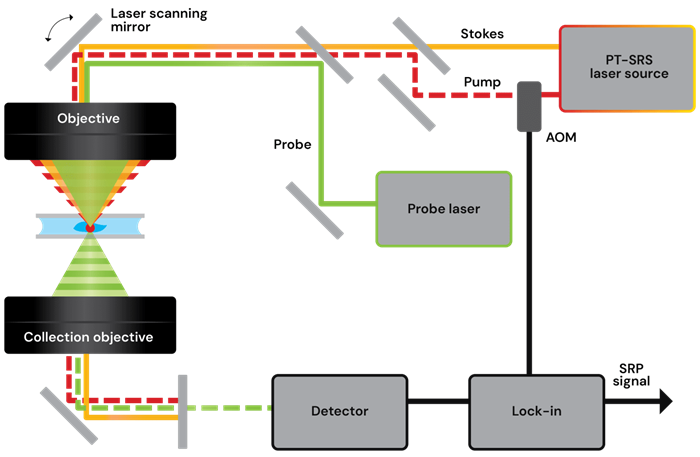
Image Credit: Photothermal Spectroscopy Corp