Raman imaging is a label-free, non-contact analytical technique that can be used to distinguish cell types and visualize subcellular structures without changing the biochemistry of a cell.
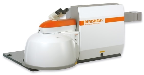
The inVia confocal Raman microscope from Renishaw provides detailed, highly specific chemical, spatial and structural information on many types of molecules at a high spatial resolution, making it ideal for live cell analysis and imaging.

Raman imaging enables cells to be identified and characterized based on their complete chemical profile and provides sub-micrometer imaging, without the need for fluorescently labelled targets.
Raman spectra reveal the overall cellular molecular composition and the heterogeneity within a cell population. The cellular molecular profile enables comparisons of different cell types and specific organelles and the fine cellular details that can be obtained mean the spatial relations of biomolecules and organelles can be visualized. The relationship between biomolecules/organelle size and distribution and cell function can also be investigated.
For live cell analysis and imaging, the inVia can be used with an incubator to allow for the control of CO2, temperature and humidity and for the study of cellular biochemical changes and responses to changing conditions.
Some of the chemical and morphological changes in cells that can be investigated include:
- Cellular uptake of drugs or drug carriersi
- DNA/RNA fragmentation and membrane blebbing during apoptosisii
- Lipid polarization in transit amplifying cellsiii
- Increases in phospholipids and vesicles during autophagyiv
- Changes in nuclear size and nucleic acid content in cancerous cells
- Metabolic changes in response to environmental stimuliv
- Increases in lipid droplets during stem cell adipogenesis
- Increased calcification during osteogenesisvi
- Redox of haem proteins in relation to cell functionvii
The 3D chemical imaging enabled by inVia provides insight into the distribution of chemical species in spaceviii. This information rich imaging also allows organelle volume to be estimated and axial data to be obtained.
The instrument is ideal for cellular studies, providing chemical data simultaneously across a wide range of molecules ranging from proteins, lipids and nucleic acids to minerals and pigments. The effects of molecular changes on biological systems can also be tracked and correlative data can be gathered with parallel techniques because samples can be reused.
The inVia features StreamHR™ imaging, providing a high spatial resolution and confocality for the scrutiny of small features. The Slalom option with StreamLine imaging also means information can be gathered from larger areas, from whole cells to sections of tissue.
Raman tissue imaging and the Renishaw inVia confocal Raman microscope
Raman imaging can be used to simultaneously determine the molecular composition and distribution of various chemical species present in tissues, at high spatial resolution and without labelling. The technique uses a laser to provide a chemical fingerprint at each point of the area being analysed. This data can then be processed to acquire a wide range of information.
The data can be used to differentiate between healthy and diseased tissue and to chemically demarcate tissue regions. Raman imaging is also an ideal pathology tool and a useful technique for use in biological researchix, x.
In the discrimination of diseased and healthy tissue, Raman imaging can be used to differentiate objectively and accurately between tissue types based on their overall chemical signatures, without the need for fluorescent or colorimetric labeling or disease maker discovery and targeting.
Chemical demarcation of tissue regions allows visualization of anatomical layers and cell typesxi; produces detailed images at sub-micrometer spatial resolution and unveils the chemical and morphological changes in different tissue samples. In terms of the zonal information that can be obtained, Raman imaging enables determination of the following:
- The complexity of tissue organization
- The size and boundaries of disease areas
- Tumor infiltration
- The relative abundance and distribution of chemical species
As a pathology tool, Raman imaging has a number of applications including the definition of tumor margins, the highly sensitive and specific discrimination of cancer stages, the identification of biochemical changes occurring with cancer formation and cancer progression and the identification of markers for early-onset diseaseix,x.
Raman imaging is also an ideal technique for use in biological research enabling researchers to study the distribution concentration, conformation, spin and redox states and orientation of biomolecules and also to compare these properties between samples. Overall, the technique allows for valuable insights into biological systems and a better understanding of them.
The inVia confocal Raman microscope
Featuring StreamHR™ imaging technology, the inVia enables high speed mapping—without tissue damage—and high confocality imaging for the scrutiny of small features. The option of StreamLine imaging with Slalom allows for a quick overview of tissue samples. Surface and LiveTrack™ enable information-rich images to be captured from uneven surfaces. The inVia also offers researchers the flexibility to switch between standard and high confocal imaging and queue up measurements mean data collection can be maximized.
References:
i) McAughtrie et al (2013) Chem Sci 4: 3566-72
ii) Uzunbajakava et al (2003) Biophys J. 84(6):3968-81
iii) Treffer et al (2012) Biochem Soc Trans 40:609-14
iv) Isabelle et al (2013) J Cancer Res Therap Oncol 1: 1-5
v) Schulze et al (2011) Applied Spectrosc 65 (12): 1380-6
vi) McManus et al (2011) Analyst 136:2471-81
vii) Armstrong et al (2015) Nature Comm 6:7405
viii) Lau et al (2014) Biomedical Spectroscopy and Imaging 3: 237-247
ix) Lau et al (2016) Proc of SPIE 27040B-3
x) Gaifulina et al (2014) Austin J Clin Pathol 1(5):3
xi) Bonifacio et al (2010) Analyst 135: 3193-3204
About Renishaw

Renishaw is a global company and a recognised leader in Raman spectroscopy. Offering high performance optical spectroscopy products for over 20 years, Renishaw’s Spectroscopy Division is passionate about manufacturing the very best Raman products. It has a team of scientists and engineers specialising in the development, application and production of high performance, configurable Raman spectrometers.
With a foundation built on sound engineering, these systems offer the highest levels of flexibility and performance and are used by academics and industrialists to tackle analytical problems across a broad range of application areas, including life science, chemicals; materials science; pharmaceuticals; semiconductors; forensics; gemmology; antiquities; and green energy, such as photovoltaics.
Renishaw’s Raman systems are easy to use and produce repeatable, reliable data, even from challenging samples. We aim to be a long-term partner, offering quality products that meet customers’ needs, both today and into the future, backed up by expert technical and commercial support.
Sponsored Content Policy: News-Medical.net publishes articles and related content that may be derived from sources where we have existing commercial relationships, provided such content adds value to the core editorial ethos of News-Medical.Net which is to educate and inform site visitors interested in medical research, science, medical devices and treatments.