In dental studies, microscopy is considered a versatile tool. While optical microscopy finds usage in day-to-day practices in dental clinics, the ability of electron microscopy to offer a large variety of surface information makes it perfect for use within a large variety of research subjects as well. The following examples offer added insights into how in-detail scanning electron microscopy (SEM) can be utilized in a variety of dental studies.
How Enamel Erosion can be Impacted by Toothpaste
A well-researched study, published by Colombo et al. [1], highlights the manner in which enamel erosion — which can occur due to the consumption of soft drinks — can be impacted by toothpaste. Colombo et al. compared three different types of enamel, by observing the enamel surface and using a SEM and morphological analysis. These three types of enamel were:
- An intact enamel surface;
- An enamel surface treated with Coca-Cola (i.e. soft drink of choice);
- An enamel surface treated with different toothpastes
It was noted that specimens treated with fluoride toothpaste displayed honey-comb structures that are characteristic of demineralized enamel. Nevertheless, they showed a healthier surface than samples treated with purely soft drinks.
After treatment, the samples that were the closest to the original/healthy state of enamel were those that were treated with Zinc-Hydroxyapatite toothpaste. Thus, Colombo et al. concluded that the SEM study enabling a qualitative understanding of the processes of demineralization of the enamel surface.
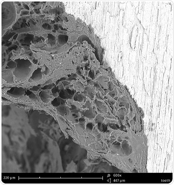
Figure 1. This is what a tooth filling interface looks like with an electron microscope
The Impact of Laser Usage in the Treatment of Root Channel
The focus of another study by Shahriari et al. [2] was the impact of laser usage in root channel treatment. Through the course of this experiment, the identification of organic/inorganic debris and smear layers on the root canal walls was done using the SEM. This was conducted after endodontic preparation, thus allowing the researchers to obtain images of dentinal tubules. From their analysis, Shahriari et al. concluded that different concentrations of sodium hypochloride activated using a laser were effective in the removal of the smear layer. However, the standard protocol still proved to be more effective than the tested one.
The Presence of Bacterial Adhesion on Sample Surfaces after Polymer Deposition
The use of scanning electron microscopy is not just apt for imaging teeth surfaces or tooth filling interactions. This technique also finds applications in tooth wear. Tupinambá et al. [3] provide insights into the usage of polymer films for conventional and self-ligating metallic brackets. These scholars performed SEM analysis to assess the presence of bacterial adhesion on sample surfaces after polymer deposition.
To look at slot and wing areas, backscattered electrons (BSE) were used. In effect, they concluded that the Hexamethyldisiloxane (HMDSO) polymer deposition on conventional brackets tended to reduce the surface roughness and the biofilm formation. Moreover, there is a need for further studies regarding polymers to evaluate the influence of self-ligating brackets.
As is evident here, a scanning electron microscope proves to be an extremely powerful tool in the production of high-quality images of surface structures, thanks to its ability to provide detailed surface information within a wide variety of research subjects.
References
[1] Remineralizing effect of a zinc-hydroxyapatite toothpaste on enamel erosion caused by soft drinks: Ultrastructural analysis, Colombo et al., J Clin Exp Dent 2017; 9 (7): e861-8.
[2] Efficacy of Sodium Hypochlorite Activated With Laser in Intracanal Smear Layer Removal: An SEM Study, Shahriari et al., J Lasers Med Sci 2017 Winter; 8 (1): 36-41.
[3] Bacterial adhesion on conventional and self-ligating metallic brackets after surface treatment with plasma-polymerized hexamethyldisiloxane, Tupinambá et al. Dental Press J Orthod 2017 July-Aug 22 (4): 77-85.
About Phenom-World
Phenom. Making SEM personal
Phenom, world’s fastest Desktop Scanning Electron Microscope takes your imaging performance to a higher level. The Phenom desktop scanning electron microscope (SEM) helps customers stay competitive in a world where critical dimensions are continuously getting smaller.
The Phenom desktop SEM combines superb imaging power up to 130,000x and outstanding technical performance with precise depth of focus and chemical contrast. It is the smart, affordable and market’s fastest solution that enables engineers, technicians, researchers and educational professionals to investigate micron and submicron structures.
The Phenom electron microscope is the ideal tool for investigating any sample and analyzing the sample composition in a short span of time. With little sample preparation needed and the incredibly short time to image, the use of the Phenom desktop SEM improves the efficiency and effectiveness of quality control and production process within high level organizations all over the world.
Phenom is known for its ease-of-use, speed and reliability.
- Phenom XL—The only desktop SEM with a 100mm x 100mm sample size and scanning area.
- Phenom ProX—The ultimate all-in-one high resolution imaging and X-ray analysis system.
- Phenom Pro—The high-end desktop SEM with superb imaging power for all markets and applications.
- Delphi microscope—The world’s first fully integrated SEM and fluorescence microscope.
- Automated Software Applications—Automated and market-specific software solutions enable Phenom users to extract maximum information from images made with the Phenom desktop SEM.
- Optimized Sample Holders—Phenom sample holders are designed for optimizing the sample acceptance and loading speed and guarantee the fastest time to image in the market.
Markets and applications
Phenom-World helps you to stay competitive in a world where critical dimensions are continuously getting smaller. The Phenom desktop SEM offers direct access to the high-resolution and high-quality imaging necessary in a large variety of applications. It is an affordable solution that enables engineers, technicians, researchers and educational professionals to visualize micron and submicron structures.
- Materials Science
- Life Sciences
- Earth Sciences
- Electronics
- Forensics
- Industrial Manufacturing
Sponsored Content Policy: News-Medical.net publishes articles and related content that may be derived from sources where we have existing commercial relationships, provided such content adds value to the core editorial ethos of News-Medical.Net which is to educate and inform site visitors interested in medical research, science, medical devices and treatments.