Introduction
Scanning electron microscopy (SEM), in the last several years, has made a major impact on different fields of biology research that are either associated with the physiology and anatomy of plants, or with the use of plant fibers in various industrial applications.
Enzymatic degumming
There is an unlimited market for fibers and fiber materials, including natural fibers such as hemp fibers. Hemp fibers are used extensively and can be acquired from Cannabis sativa L, which can easily grow as high as 4 meters.
They are resistant and long-lasting, with their diameters ranging between 16 and 50 micrometers. Hemp fibers have been used for many years for making papers, ropes, clothes, and canvas. Lately, these fibers have been used in the automobile industry to strengthen paint.
Conversely, the hemp phloem also contains wax, pectin, lignin¸ and hemicelluloses in addition to other impurities that are generally eliminated through the degumming process1. While there are a number of degumming methods, chemical degumming is traditionally used because hemp is highly resistant to water.
However, this method can consume plenty of energy and water, which presents serious environmental concerns. As a result, a research team has developed a novel technique on the basis of enzymatic degumming; the method is effective and removes the above-mentioned problems.
In order to find out how enzymatic degumming affects the quality of hemp fibers, a combination of SEM and fluorescent microscopy was used to view both untreated and treated hemp fibers. The objective of this analysis was to determine the content and quality of hemp fibers in proteins and judge the efficiency of the enzymatic degumming process.
The study could show that this tested biochemical combined degumming process provides a suitable solution in terms of environmental and economical impact and hence, can be easily industrialized.
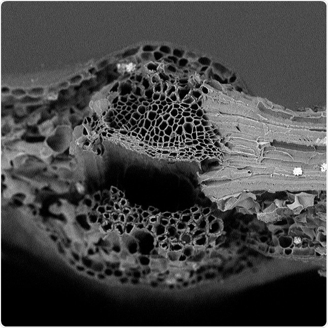
Figure 1. Imaging an ivy leaf with SEM
SEM and correlative light and electron microscopy (CLEM)
In this case, the application of correlative light and electron microscopy (CLEM) may have been advantageous, because with this method both the fiber’s morphology and the fiber’s richness in unwanted proteins can be visualized simultaneously.
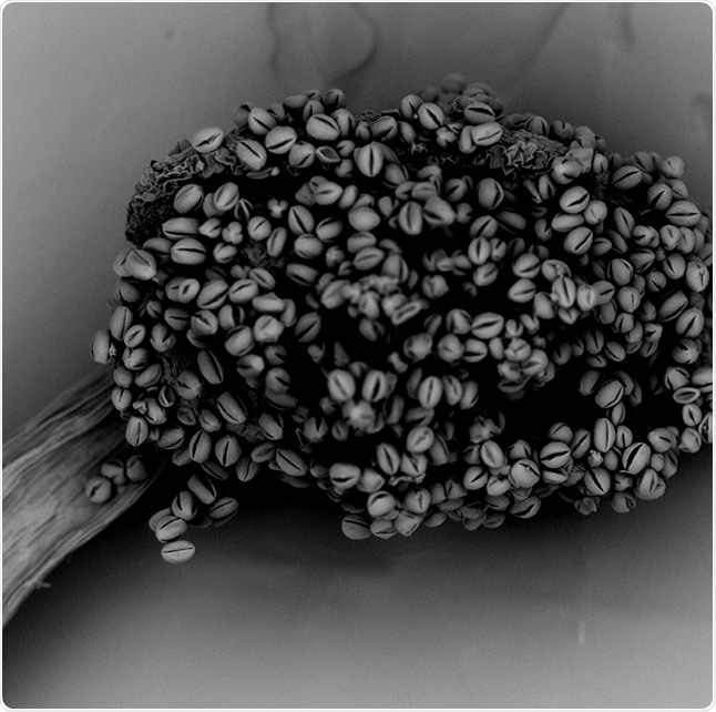
Figure 2. Plant stamina examined with SEM
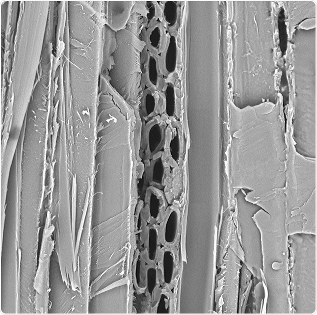
Figure 3. Example of wood images with SEM
Moreover, SEM can be useful in environmental studies. For instance, the existing political situation in Nigeria resulted in abundant oil spills in the ocean, which considerably damaged the environment and also led to significant wastage of natural resources.
This problem triggered a great deal of interest among the researchers who went on to study the surface of a plant called Delonis regia2. This plant can absorb oil in the ocean but at the same, it also releases some of this oil under particular conditions.
The researchers attempted to boost the percentage of oil which they recovered from the plant following its absorption of crude oil, and discovered that oil percentage can be increased by reducing the hydroxyl groups on its surface.
Untreated, treated, and oil covered samples of Delonis regia were compared using the SEM. The technique also made it possible to visualize surface modifications, which resulted in better absorption and diffusion of oil.
Conclusion
The SEM serves as a robust tool for analyzing the morphology of plant fibers as well as to evaluate their reaction and quality in certain conditions. Thus, the images acquired with SEM present a strong platform for various studies and help achieve desirable outcomes.
References
- An Efficient Method of Bio-Chemical Combined Treatment for Obtaining High-Quality Hemp Fiber. Fang et al. Bioresources (2017)
- Kinetic studies of surface modification of lignocellulosic Delonix regia pods as sorbent for crude oil spill in water. Onwuka et al. Journal of Applied Research and Technology (2016)
About Phenom-World

Phenom-World believe breakthroughs happen when complex nanotechnology is made intuitive, easier to use and brought within reach. As the leading global supplier of desktop scanning electron microscopes, their aim is to make imaging and analysis at the nanoscale available to every scientist in every lab.
That’s why Phenom-World invest their time and effort into developing high-quality electron microscope solutions that are functionally rich, yet simple to use. Because their mission is to create technology that has a real impact on how people work.
This takes innovative thinking, collaborative working and an entrepreneurial attitude. At their home in the high-tech region of Eindhoven in The Netherlands, Phenom-World have a group of passionate people who are given the freedom they need to innovate. Plus a highly skilled team of specialists in more than 40 countries who can provide support on a global scale.
Cooperation and co-creation are key to Phenom-World's success. They work closely with their partners to ensure they have the latest technologies, state-of-the-art equipment and applications for imaging and analysis. So they can get fast and accurate results and achieve more in nanotechnology.
Phenom-World is globally the yearly number 1 manufacturer of desktop scanning electron microscopes and imaging and analysis packages.
Sponsored Content Policy: News-Medical.net publishes articles and related content that may be derived from sources where we have existing commercial relationships, provided such content adds value to the core editorial ethos of News-Medical.Net which is to educate and inform site visitors interested in medical research, science, medical devices and treatments.