The microbes found in farm, zoo, pet animals as well as in the environment (water, soil, air) that make human beings fall sick, irrespective of the infection path, are referred to as foodborne pathogens. Around 250 different types of viruses, bacteria, algae, parasites, and molds constitute the family of foodborne microbes. All of them are so minute that they cannot be seen without a microscope.
Although light microscopy makes it possible to observe most species, electron microscopy allows detailed checking of the morphology of foodborne pathogens. Therefore a more detailed observation of the interactions between human cells and bacteria or inter-bacterial interactions is possible through electron microscopy. Such details are necessary to understand the pathogen and help develop potential cures.
These are some of the most common bacterial pathogens, which can cause foodborne diseases and result in more dangerous complications. These include:
- Salmonella enterica
- Clostridium perfringens
- Campylobacter jejuni
- Shigella spp
- Listeria monocytogenes
Salmonella enterica
A rod-shaped bacterium Salmonella enterica infects humans and has a wide range of other animal hosts. The most common hosts are cattle and poultry, but domestic pets such as cats and hamsters can also be infected by these bacteria. Pasteurization and proper cooking of food before consumption removes these pathogens. Salmonella enterica causes abdominal cramps, diarrhea, fever, and vomiting for 12 to 72 hours after infection.
Clostridium perfringens
Clostridium perfringens is another major cause of food poisoning, where symptoms appear more rapidly (typically within six hours of the infection) than with Salmonella. This bacterium is found in poorly cooked meat and poultry, but is also found in well-cooked but improperly-stored food. The symptoms of this infection are fever, diarrhea, abdominal cramps, and vomiting. If left untreated, food poisoning could cause the C. perfringens to settle in the intestine allowing it to secrete a necrotic toxin, which can result in fatal intestinal hemorrhage.
Campylobacter jejuni
Campylobacteriosis causes an inflammatory, often bloody, diarrhea or dysentery accompanied by abdominal cramps and fever. Campylobacter jejuni is a comma-shaped, spiral bacterium that is commonly found in unpasteurized milk and poultry. The pathogen is also transmitted by fecal-oral route or by sexual contact.
Shigella spp
Shigella is a bacterium commonly found in humans and gorillas. It is transmitted through uncooked food or poor hygiene and causes Shigellosis. This infection can be prevented by thorough food cooking and frequent hand washing. It is characterized by delayed symptoms, which can last for weeks. Some of the most common symptoms are painful bowel movements, vomiting, abdominal pain, and severe diarrhea.
Listeria monocytogenes
Listeria monocytogenes is a rod-shaped bacterium that causes infections of the central nervous system along with Gastroenteritis. It is present everywhere and is resistant to extremely cold temperatures and hence, this infection cannot be prevented by storing food in the refrigerator. Diarrhea and abdominal pain are the main symptoms. Two common bacterial images seen through scanning electron microscopy are shown below:
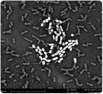
Figure 1. SEM image of E-Coli bacteria.
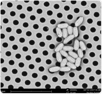
Figure 2. SEM image of Bacillus Pumilus.
Fungal Infections
Unlike fungi, which is commonly known as mold, bacterial infection of food is not usually visible to the naked eye. Several thousands of known species of mold can grow on clothes, carpets, leather, and wood. However, most significantly, molds can grow on food if placed in moist conditions. Several common types of mold reproduce by making spores. When these spores settle down on a moist food source, they germinate and form a branching network of cells called hyphae, which are visible. The impact on human health depends on the nature of the fungi and the length of exposure.
The two figures shown below represent Aspergillus niger and Rhizopus stolonifer, isolated from coffee and bread, respectively. Aspergillus niger is a common food contaminant that is also known as black mold because it is identified by its black spores, even though the contaminant can be confused with other species. A. niger rarely causes human diseases when compared to other Aspergillus species, but if present in large quantities, it can cause fungal ear infections.
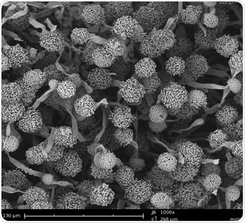
Figure 3. SEM image of Aspergillus niger spores found in coffee.
Rhizopus stolonifer is also known as Black Bread Mold. Resembling threads, this bacterium consumes nutrients from bread and damages the surface of its host. R. stolonifer is usually found in cosmopolitan locations, and can cause opportunistic infections of humans as well as infection in vegetables and fruits.
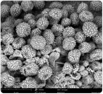
Figure 4. SEM image of Rhizopus stolonifer collected from bread.

This information has been sourced, reviewed and adapted from materials provided by Phenom-World BV.
For more information on this source, please visit Phenom-World BV.