Fragment-based drug design (FBDD) has led to a revolution in contemporary drug discovery, facilitating a bottom-up approach to the identification and evolution of bioactive small molecules into viable drug candidates. FBDD has thus far resulted in seven FDA-approved drugs and more than 50 clinical trials.
NMR spectroscopy is considered to be the gold standard method for fragment screening. In this context, fragment screening can be understood as the identification of a protein’s interaction with small molecules of molecular mass below 300 g/mol.
NMR fragment screening had previously necessitated the use of a high-field NMR spectrometer, but recent developments by NexMR have enabled the screening of fragment libraries on a cryogen-free benchtop NMR spectrometer; for example, Oxford Instruments’ X-Pulse.
NexMR’s technology illuminates samples to better enhance the NMR signal of photoinducible fragments, allowing these to be detected within seconds to minutes.
This improved sensitivity is realized by spiking the sample with a photosensitizer and an oxygen-quenching enzyme cocktail. The sample is illuminated for several seconds while sitting in the magnet.
Photo-induced signal enhancement is the most rapid means of improving NMR instruments’ sensitivity. It can also be performed at room temperature in an aqueous buffer - a highly preferable condition for drug discovery.
Fragment screening requires that sample illumination be compatible with an automated sample changer. This ensures high enough throughput to take full advantage of the ultrafast measurement capabilities of NexMR’s photoinducible fragment libraries.
Standard benchtop NMR setups are unsuited to illumination via automated sample changers. Manual intervention is required because the light coupling requires a physical connection between the light source and the sample via a waveguide.
A customized version of the X-Pulse is available, which features integrated light capability alongside an automated setup process. This setup can accommodate a screening rate of one sample every five minutes on a benchtop NMR spectrometer, the equivalent of 288 samples per day.
Oxford Instruments’ X-Pulse is a modular broadband benchtop spectrometer that offers unparalleled flexibility to virtually any analytical or research laboratory. As well as its capacity to accommodate a high-throughput autosampler, the X-Pulse can also be equipped with a flow cell or variable temperature (0 °C to 65 °C).
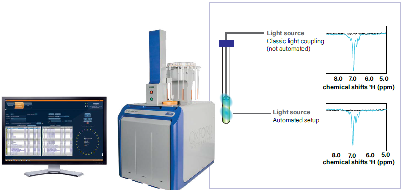
Figure 1. Automated X-pulse with light and measurement of the signal-to-noise. Image Credit: Oxford Instruments
Performance of NexMR’s fragment kits with X-Pulse
NexMR’s fragment libraries (NMhare) contain a range of aromatic scaffolds, all of which can be observed using an X-Pulse benchtop NMR spectrometer with integrated light capability.
Following a few seconds of illumination, low concentrations of the fragments in an aqueous buffer (200 µmol/ℓ) can be detected within a single two-second scan (Figure 2).
In the example presented here, a total of 16 of the 18 tested fragments demonstrated a notable signal-to-noise ratio (SNR) following a single scan at 200 µmol/ℓ.
The median SNR was determined to be seven (with an average of 20), while the maximum observed SNR with a single scan was 112, and the minimum was two.
It was noted that an accumulation of 16 scans results in a minimum observable SNR of 10, allowing for quantitative peak analysis. The maximum SNR was determined to be 450; with a median of 30, and an average of 82.
Without photo-boosted NMR, these types of experiments would take more than 100 hours, but this level of performance enables fragment screening within just two minutes.
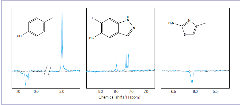
Figure 2. Photo-boosted NMR spectra (blue) of three fragments with diverse aromatic scaffolds after 2 seconds of illumination and the same experiment without illumination (black). Image Credit: Oxford Instruments
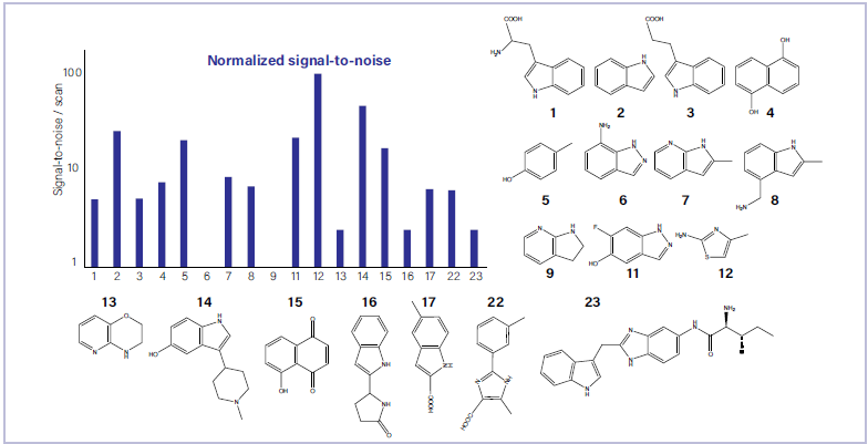
Figure 3. Signal-to-noise ratio (SNR) per molecule and scan. Each molecule was measured at a concentration of 200 μmol/ℓ. Image Credit: Oxford Instruments
Low micromolar measurements
For quantitative peak analysis, an SNR of 10 is typically required. Ligand-observed NMR screening is performed at 50-200 μmol/ℓ ligand concentrations at high magnetic fields.
In the example presented here, SNR was evaluated at different concentrations for tryptophan and cresol - two emblematic molecules yielding photo-boosted NMR.
Sixteen scans were performed in two minutes, confirming that it was possible to observe down to 20 μmol/ℓ for each molecule (Figure 4). For quantitative analysis, it is advisable to use 64 (eight-minute) scans for 20 μmol/ℓ screening and 16 scans for 50 μmol/ℓ screening.
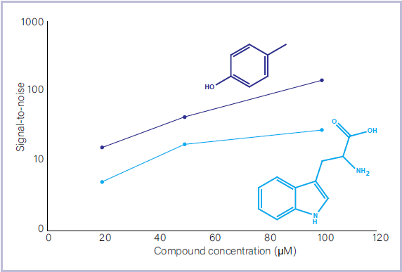
Figure 4. Measurement of photoinducible reference fragments at different micromolar concentrations. Image Credit: Oxford Instruments
Fragment screening with the X-Pulse benchtop NMR spectrometer and NMhare photo-inducible libraries
Small molecules have the potential to interact with larger macromolecules, forming a complex in a slow rotational motion. Under these circumstances, small molecule polarization is transferred to the protein via the nuclear Overhauser effect (NOE). This results in a reduction in signal intensity.
Identifying a protein-fragment binding event is possible by tracking the signal reduction after a protein of interest has been added. This fragment screening technology has been documented previously by NexMR and the Riek Lab (ETH Zürich).1 The signal reduction can be quantified with the polarization ratio (PR). This is defined as:

In the formula shown here, IPL and IL refer to the integral of the hyperpolarized ligand peak with and without protein, respectively.
The molecules here were tested against known protein targets to observe the signal reduction. This was done by binding all molecules that exhibited photo-boosted signals.
One fragment was considered a ‘hit,’ because this showed an especially significant reduction in the signal. It was noted that the hit was seen for the PIN1 protein, but the KRAS protein exhibited a characteristic decrease in signal.
Should PIN1 have been a druggable target protein, medicinal chemists would likely begin to base their drug design around this fragment because it had been shown to bind with the target protein.
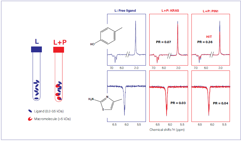
Figure 5. Fragment screening of two fragment molecules against KRAS and PIN1, two oncoproteins. The polarization ratios (PR) are shown to reflect quantitative analysis. Image Credit: Oxford Instruments
Summary
This article highlighted the potential of NexMR’s fragment library and Oxford Instruments’ X-Pulse with autosampler to enable automated high-throughput fragment screening.
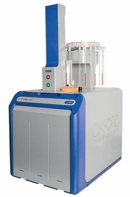
Image Credit: Oxford Instruments
Using photo-induced signal boosting, the experiment time was reduced from over 100 hours to two minutes. This significant reduction enabled the testing of fragments for their photo response and subsequent screening of protein molecules for binding, all within a single working day.
This highly reliable, high-throughput screening approach can enable academic and industrial pharmaceutical researchers to accelerate drug development in their laboratories while leveraging the many benefits of small-footprint, cryogen-free benchtop NMR.
References and further reading
- F. Torres et al., J. Am. Chem. Soc., 2023, 145, 12066-12080.
Acknowledgments
Produced from materials originally authored by Oxford Instruments.
About Oxford Instruments
Oxford Instruments is a leading provider of high-technology tools and systems for research and industry, dedicated to accelerating breakthroughs that create a brighter future for our world. With a global presence, we are committed to innovation and excellence, offering cutting-edge solutions that enable researchers and industry professionals to achieve breakthroughs in their fields. Our advanced technologies deliver numerous benefits through unparalleled precision and reliability, allowing users to obtain accurate and reproducible results. By utilising Oxford Instruments' innovative solutions, research is accelerated, productivity is enhanced, and innovation is achieved in various fields, including materials analysis, life sciences, semiconductors, physics, chemistry, and food sciences. We take pride in being a trusted partner for those aiming to push the boundaries of scientific and industrial advancements, providing the tools and support necessary to realise their visions.
Atomic force microscopy: These advanced instruments are utilised for high-resolution imaging and precise measurement of surface properties at the nanoscale level. They offer detailed topographical information and enable accurate measurements of features such as height, roughness, and mechanical properties.
Light microscopy: Our solutions encompass a comprehensive range of advanced imaging systems that utilise visible light for examining samples at the microscale. Equipped with high-quality lenses, cameras, and illumination systems, these optical microscopes deliver detailed images with exceptional clarity and resolution. They find applications in fields such as biology, materials science, and forensics.
Deposition and etching: Our advanced tools are specifically designed for the precise fabrication and modification of materials at the nanoscale. They include a variety of deposition techniques like physical vapor deposition (PVD), chemical vapour deposition (CVD), and atomic layer deposition (ALD), as well as etching processes, such as plasma etching and reactive ion etching (RIE). These tools empower users to create customised materials and devices with exceptional control and precision.
Electron microscopy analysis: Our high-performance tools are tailored for materials characterisation, particle analysis, and sample manipulation at the nanometre scale. Techniques such as Backscatter Electron and X-ray (BEX), Energy Dispersive Spectroscopy (EDS), and Electron Backscatter Diffraction (EBSD) enable imaging, chemical analysis, and crystallographic characterisation of materials at atomic and nanoscale levels.
Optical imaging and spectroscopy: Our optical imaging solutions comprise a range of advanced technologies and systems for capturing and analysing images using light. From state-of-the-art optical microscopes to spectroscopy systems and imaging software, these solutions provide high-quality images and data for applications such as materials characterisation, biological research, and semiconductor analysis.
Nanoindentation: Our high-resolution, MEMS-based nanoindenters are designed to measure the mechanical properties of materials, including hardness, elastic modulus, stiffness, and creep behaviour. These instruments offer superior fabrication tolerances, enhancing sensitivity, resolution, and repeatability beyond conventional technology limits.
Nuclear magnetic resonance: We offer a variety of benchtop Nuclear Magnetic Resonance (NMR) instruments for research, industrial quality assurance/control, and rock core analysis. These instruments provide advanced capabilities for chemical analysis, materials characterisation, and more.
Raman microscopy: Our advanced imaging systems are tailored for high-resolution microscopy and analysis, offering cutting-edge technology and precision optics for detailed insights into samples at the micro- and nanoscales. Trusted by researchers and scientists in fields such as materials science, life sciences, and nanotechnology, these systems are available in various models to suit different applications.
X-ray technologies: Our technologies encompass a range of advanced systems and solutions for materials analysis and characterisation. With cutting-edge capabilities and high sensitivity, Oxford Instruments X-ray technologies are widely used in various industries and research academic institutions for applications such as material identification, quality control, and research in fields like geology, metallurgy, and semiconductor manufacturing.
Sponsored Content Policy: News-Medical.net publishes articles and related content that may be derived from sources where we have existing commercial relationships, provided such content adds value to the core editorial ethos of News-Medical.Net which is to educate and inform site visitors interested in medical research, science, medical devices and treatments.