Sponsored Content by Bruker OpticsReviewed by Olivia FrostNov 5 2026
Can microplastics permeate biological boundaries and build up within internal organs? In biomedical and environmental studies, this has become one of the most crucial questions. Specific detection within organs remains analytically challenging, even though evidence of microplastics in digestive systems is well documented.
The proposition that small enough polyethylene (PE) microplastics can pass the intestinal barrier and build up in liver tissue was studied by a university research group. This article shares the analytical method employed to collect chemically specific evidence of PE particles inside the tissue.
As an experimental model, cryo-sectioned samples of chicken liver were prepared. Using optical microscopy, small dark debris was observed within the tissue (Figure 1).
Raman microscopy was employed to classify possible PE particles from biological structures, as optical contrast and morphology alone were unable to determine their chemical composition.
Characterization of fine particles using Raman microscopy
Infrared-based analysis did not give strong confirmation of polyethylene within the liver tissue even though it is the routine approach. The particles under question were challenging to resolve through the use of infrared microscopy as they were only an estimated 1–5 µm in size.
As its high spatial resolution covers the chemical characterization of particles within this size threshold, Raman microscopy was chosen. This characteristic makes Raman spectroscopy ideal for uncovering the smallest microplastic particles residing in biological tissue.
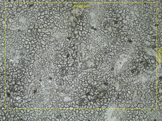
Figure 1. Microscopic image of a chicken liver cryo-section. Dark spots are suspected to be microplastic particles. Image Credit: Bruker Optics
Results
The analyzed region spans an estimated 491 × 345 µm, with a 1.2 µm spatial sampling resolution, producing a dataset capable of resolving micron-scale particles inside the tissue matrix.
To ensure full coverage, a cumulative acquisition time of 15 hours (Figure 2, top right) was planned for a standard grid-based Raman mapping.
Random Scanning was employed to eliminate spending measurement time on tissue areas that lacked signals of interest, because the target particles were presumed to be unevenly distributed and sparse.
By selectively measuring locations inside the defined area that were rich in chemical information, this strategy increased the probability of locating polyethylene particles while lowering the time taken to achieve relevant results.
The instrument had already detected and analyzed the majority of the polyethylene particles (Figure 2, top left) within the first three to four hours. Even at this early stage, the corresponding Raman image closely matched the end dataset in both statistical significance and spatial distribution.
The distinctive Raman signature of polyethylene was detected at each labeled pixel. The respective complete spectra (Figure 2, bottom) validated the particles’ chemical identity, offering clear proof of polyethylene microplastics present in the liver tissue.
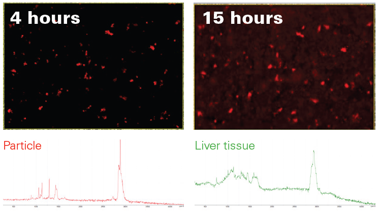
Figure 2. Comparison of the Raman image acquired with Random Scanning after four hours (top left) with the complete 15-hour Raman image (top right). Clearly, most particles were found by the algorithm. Found particles were unambiguously identified as PE (bottom left) and clearly stand out from the liver tissue matrix (bottom right). Image Credit: Bruker Optics
Conclusion
This investigation effectively demonstrated the direct identification and detection of polyethylene microplastics in liver tissue by using intelligent Random Scanning with confocal Raman microscopy.
Raman microscopy is especially appropriate for the measurement of very small particles in intricate biological backgrounds, where high spatial resolution is crucial. It delivers a valuable complementary method for studies that go further than the resolution limits of infrared approaches.
However, infrared imaging continues to be a foundation of microplastics studies due to its unrivaled rapid screening capacity and fast measurement times.
Acknowledgments
Produced from materials originally authored by Dr. Yan Di from Bruker Optics GmbH & Co. KG.
About Bruker Optics
Bruker Optics, part of the Bruker Corporation, is one of the world’s leading manufacturers and worldwide suppliers of Fourier Transform Infrared, Near Infrared and Raman spectrometers.
Bruker entered the field of FTIR spectroscopy in 1974. The early instruments set new standards in research FTIR with evacuable optics, high resolution, and automatic range change. Since then, the product line has been continuously expanding with instruments suitable for both analytical and research applications with exceptional performance characteristics.
Today, Bruker Optics offers complete technical solutions for various markets that cover a broad range of applications in all fields of research and development as well as industrial production processes for the purpose of ensuring quality and process reliability.
Bruker Optics’ R&D and manufacturing center is located in Ettlingen, Germany; technical support centers and sales offices are located throughout Europe, North and South America, Asia, India, the Middle East, and Africa.
Sponsored Content Policy: News-Medical.net publishes articles and related content that may be derived from sources where we have existing commercial relationships, provided such content adds value to the core editorial ethos of News-Medical.net, which is to educate and inform site visitors interested in medical research, science, medical devices and treatments.