Life science microscopy plays an important role in helping us understand the complex structures, processes, and interactions that make up biological systems. From exploring molecules to diagnosing diseases, advances in imaging technologies are giving researchers a closer look at how biological systems work, revealing new insights and helping us better understand disease.
With so much happening in this field, we've brought together some of the latest research, technologies, and applications in our Life Science Microscopy Industry Focus eBook. It explores how advances in microscopy are opening new possibilities for biological discovery, supporting clinical research, and improving how scientists study and analyze biological samples.
Download the eBook
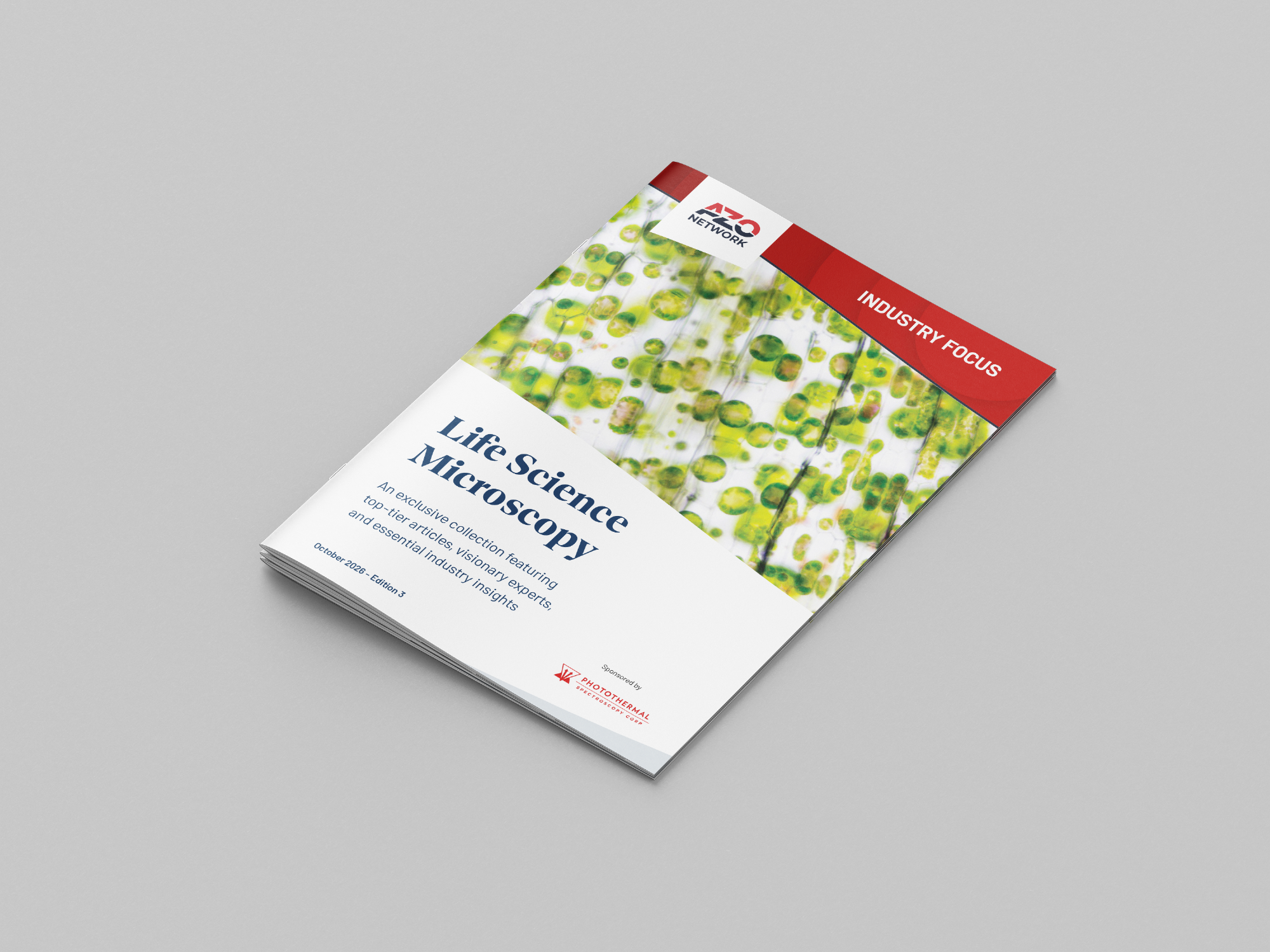
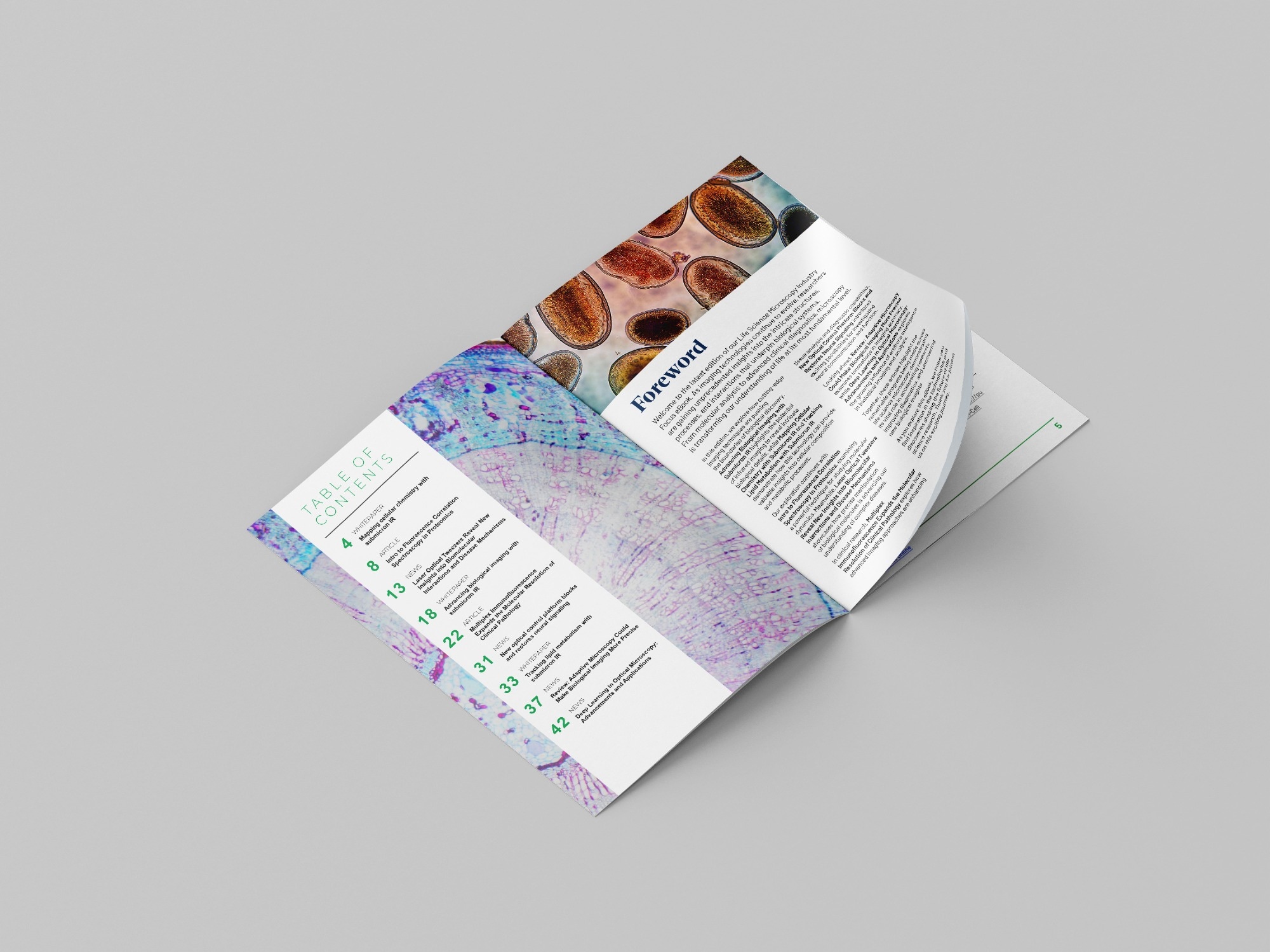
Here's a look at what you'll find inside:
- How submicron infrared (IR) imaging is revealing intricate biological structures and finer details
- How researchers are using submicron IR to map cellular chemistry and explore lipid metabolism
- The role of fluorescence correlation spectroscopy in understanding molecular dynamics and proteomics
- How laser optical tweezers are helping researchers investigate biomolecular interactions and disease mechanisms
- The latest advances in multiplex immunofluorescence and what they mean for tissue analysis and clinical pathology
- How new optical control technologies are helping scientists investigate and restore neural signaling
- The potential of adaptive microscopy to improve imaging precision in biological research
- How deep learning is becoming an increasingly valuable tool in optical microscopy and biological image analysis
Together, these topics offer a closer look at exciting developments in life science microscopy, highlighting both the technologies behind them and their practical applications in biological research.
Whether you work in molecular biology, cellular research, clinical pathology, proteomics, or biological imaging, this eBook is a great way to catch up on the latest developments and explore the techniques shaping the future of life science research.
Download the eBook
About Photothermal Spectroscopy Corp 
Photothermal Spectroscopy Corp (PSC) is a Santa Barbara, CA-based scientific equipment company that pioneered sub-micron IR microscopy and spectroscopy. PSC’s vision is to enable the power of sub-micron IR spectroscopy to be applied to high-value problems in both industry and academia via the adoption of O-PTIR and co-located multi-modal techniques.
O-PTIR is one of the highest growth IR microscopy techniques and addresses application areas in life science, microplastics, bioplastics, semiconductors, failure analysis, and chemical and polymeric materials.
mIRage-HSi IR multimodal microscope
mIRage-HSi combines sub-500 nm O-PTIR chemical imaging with laser-scanning speed, revealing label-free distributions of key macromolecules like lipids, proteins and nucleic acids while also resolving micro- and nanoplastic particles in complex biological samples.
Hyperspectral images are acquired in minutes and targeted single-wavenumber images in seconds, making detailed cellular studies and high-throughput particle workflows practical.
The mIRage-HSi supports multi-modal capabilities with simultaneous raman, co-located fluorescence microscopy and optical microscopy making it a powerful instrument for high resolution life science research.
Optical Photothermal IR spectroscopy
Multimodal Optical Photothermal infrared spectroscopy (O-PTIR) used on the mIRage microscope platforms is a result of nearly 2 decades of expertise in photothermal based IR spectroscopy.
O-PTIR: How it works
O-PTIR overcomes the IR diffraction limit by combining a mid-IR pulsed, tunable laser that heats the sample. When the IR laser is at a wavelength that excites a molecular vibration in the sample, absorption occurs, thereby creating photothermal effects including photothermal expansion. A visible probe laser, focused to 0.5 µm spot size, measures the photothermal response via the scattered light, as shown in the illustration above.
The component of the reflected visible laser signal that is modulated at the IR pump laser repetition rate is directly proportional to the absorption coefficient of the sample at that wavenumber. The IR pump laser can be tuned through the entire fingerprint region in one second or less, to obtain an IR spectrum.
By operating in reflection mode, O-PTIR eliminates several longstanding limitations and has substantial benefits for the IR community, including sub-micron resolution using a non-contact optical method. The sub-micron resolution is demonstrated (right), showing reflection mode spectra on a multi-layer packaging film measured 0.5 µm apart with highly differentiated chemical fingerprints indicating different materials.
Measurements are collected quickly and easily without need for sample contact, unlike ATR spectroscopy. Additionally, O-PTIR provides spectra comparable to FTIR without the dispersive artifacts observed in ATR. By operating in reflection mode, the need for thin samples is also eliminated, leading to dramatically easier sample preparation and faster turnaround times.
Sponsored Content Policy: News-Medical.net publishes articles and related content that may be derived from sources where we have existing commercial relationships, provided such content adds value to the core editorial ethos of News-Medical.net, which is to educate and inform site visitors interested in medical research, science, medical devices and treatments.