Sponsored Content by Bruker OpticsReviewed by Olivia FrostNov 5 2026
Mussels are widely trusted sentinel organisms for tracking coastal microplastics. Their tissue load mirrors local contamination over time because they are suspension feeders that filter significant amounts of water and gather particles from the nearby water column.
As opposed to visual observation alone, chemical analysis is required to determine the polymer class of the collected particles. Particles obtained from digested tissue cover a large area and are present alongside organic and mineral matter that cannot be identified from plastics visually.
FTIR (Fourier-Transform Infrared) imaging can create spatially resolved chemical maps of large filter areas and even entire filters, identifying each particle by its infrared spectrum and providing morphology and size information in a single assessment.
This article outlines its deployment for the characterization of microplastics in two species of mussels, sampled across two seasons, from Guanabara Bay, Rio de Janeiro, as outlined by UFF Brazil’s Dr. C. Rodrigues and Dr. J. Paulo Felizardo.
Experimental
The Rio–Niterói Bridge and Jurujuba areas are two aquaculture sites in Guanabara Bay from which a total of 120 mussel individuals were collected. The Asian green mussel Perna viridis and the brown mussel Perna perna were the two sampled species.
To account for seasonal differences in particle load, collections were conducted during the rainy season (February 2025) and the dry season (August 2025).
A 10% KOH solution at a solution-to-sample volume ratio of 5:1 was used to undertake sample preparation by alkaline digestion. For 72 hours, samples were incubated at 40 °C. A 25 µm stainless steel mesh was used to filter the digested material, and the retained amount was deposited into glass beakers.
Ten milliliters of NaI solution (4.4 mol/L; 1.8 g/cm3) was added to perform density separation after 15 minutes of sonication and 15 minutes of mechanical agitation at minimum. A further 10 mL of NaI solution was introduced during agitation. This process was followed twice.
For around two minutes, the samples were then centrifuged at 500 × g, with this process repeated if suspended material could still be seen. Lastly, the low-density supernatant was vacuum-filtered onto 0.2 µm anodisc aluminum oxide filters.
A procedural blank was maintained throughout all preparation stages to verify the sample preparation procedure, excluding biological tissue.
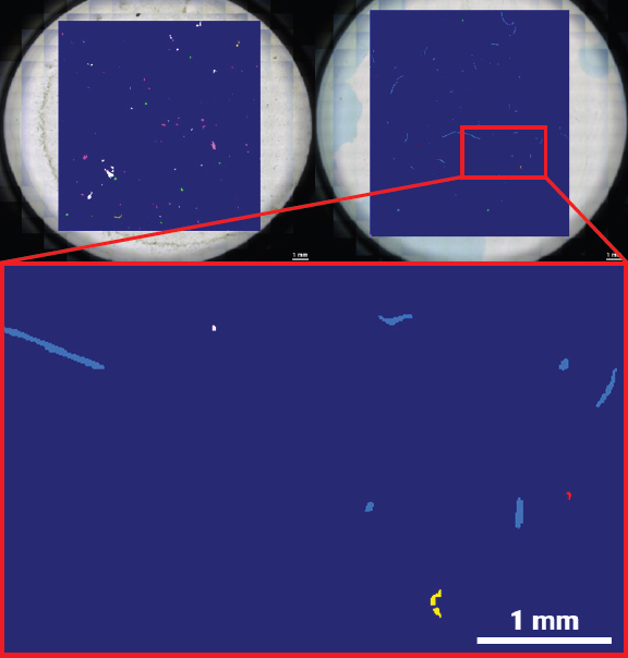
Figure 1. Data obtained from a Guanabara Bay mussel tissue sample. Left: Transmitted-light mosaic of the anodisc filter following alkaline digestion and density-based extraction. Right: Corresponding hyperspectral μFTIR map acquired with the Bruker LUMOS II and evaluated with the Bruker MPID software. Image Credit: Dr. J. Paulo Felizardo and Dr. C. Rodrigues.
Results
The Bruker LUMOS II FTIR imaging microscope was used to measure anodisc filters, and the data was analyzed using the Microplastic Identifier (MPID) software, which deploys machine learning to classify and detect particles.
Large-scale particle exposure in Guanabara Bay was deemed likely, as microplastic particles were identified in all 120 individual samples inside the measured area using the chosen preparation and µFTIR imaging procedure. A 100% occurrence rate indicates the severity of particulate pollution in Guanabara Bay and the precision of the analytical technique.
The results demonstrated that P. perna had a mean of 2.2 particles per individual, while P. viridis had a mean of 5.4 particles per mussel.
The relationship reversed when normalized to soft tissue mass, with P. viridis showing 0.19 ± 0.13 particles/g compared to P. perna’s much higher contamination level: 0.54 ± 0.45 particles/g.
This variation should be understood in the context of environmental and biological covariates, as they could reflect tissue composition, species-specific body mass, site-related exposure trends, or filtration behavior.
The primary synthetic polymer in both species of mussel was polypropylene (PP). PP had a mean length of 35.77 µm and a width of 4.33 µm, consisting mostly of fragments (92%) in P. perna.
Its pervasiveness is consistent with the low density of PP and its common applications in ropes, packaging, and other marine-related materials, but the determination of the polymer alone does not confirm source attribution.
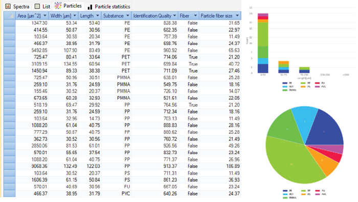
Figure 2. Exemplary MPID result obtained from a Guanabara Bay mussel tissue sample. Extensive particle information is given together with an interactive classification map. When a particle or particle class is selected, all respective information is directly displayed. Image Credit: Bruker Optics
Conclusion
FTIR microspectroscopy is ideal for the analysis of microplastics in digested biological tissues, providing chemically specific, automated polymer identification that reduces operator variability and detects particles below the visual-sorting size threshold.
The prevalence of contamination in Guanabara Bay is evidenced by the presence of microplastics in all mussel samples. Variations in mass-normalized burdens between P. viridis and P. perna suggest that species-specific biological elements must be included in standardized biomonitoring procedures.
Mussels, as filter feeders, collect microplastics from surrounding water and can serve as bioindicators of environmental contamination. Their role in seafood consumption and marine food chains also provides a potential pathway for exposure to higher trophic levels.
Continual observation is critical to evaluate possible human exposure and ecological contamination.
Acknowledgments
Produced from materials originally authored by João Paulo Felizardo (PhD), Camila Rodrigues e Silva (PhD), and Alexander Staub (PhD) from Federal Fluminense University and Bruker Optics GmbH & Co. KG.
About Bruker Optics
Bruker Optics, part of the Bruker Corporation, is one of the world’s leading manufacturers and worldwide suppliers of Fourier Transform Infrared, Near Infrared and Raman spectrometers.
Bruker entered the field of FTIR spectroscopy in 1974. The early instruments set new standards in research FTIR with evacuable optics, high resolution, and automatic range change. Since then, the product line has been continuously expanding with instruments suitable for both analytical and research applications with exceptional performance characteristics.
Today, Bruker Optics offers complete technical solutions for various markets that cover a broad range of applications in all fields of research and development as well as industrial production processes for the purpose of ensuring quality and process reliability.
Bruker Optics’ R&D and manufacturing center is located in Ettlingen, Germany; technical support centers and sales offices are located throughout Europe, North and South America, Asia, India, the Middle East, and Africa.
Sponsored Content Policy: News-Medical.net publishes articles and related content that may be derived from sources where we have existing commercial relationships, provided such content adds value to the core editorial ethos of News-Medical.net, which is to educate and inform site visitors interested in medical research, science, medical devices and treatments.