Yokogawa’s CSU-W1 Confocal Scanner sets the industry standard for live cell imaging, offering unparalleled flexibility and a four-times-wider field of view than conventional models. With three camera configurations, two pinhole size options, near-infrared observation, and an external light path, the CSU-W1 adapts to diverse research needs. Its fully motorized switching mechanisms ensure seamless automation, making it ideal for a wide range of experiments. Together, these features deliver Yokogawa’s clearest and most expansive confocal imaging experience.
Microlensed Dual Disk Design For Better Images
A Nipkow spinning disk containing about 20,000 pinholes and a second spinning disk containing the same number of microlens to focus excitation laser light into each corresponding pinhole are mechanically fixed with a motor, and very rapidly raster scan the field of view with about 1,000 laser beams when rotated. The pinhole and microlens pattern are arranged in our proprietary design to optimize raster scan. Multi-beam scanning with the CSU-W1 not only increases scanning speed, but also results in significantly lower photobleaching and phototoxicity, because multiple excitation needs only a low level of laser power at the specimen to fully excite fluorescence.
Key Features
4x Wider FOV compared to conventional models
Fully Motorized Switching for automated experiments
Multiple Configurations to suit diverse applications
Selectable Pinhole Sizes for optimized imaging
Wide
The largest FOV confocal offers a four-fold broader field of view than the standard model.
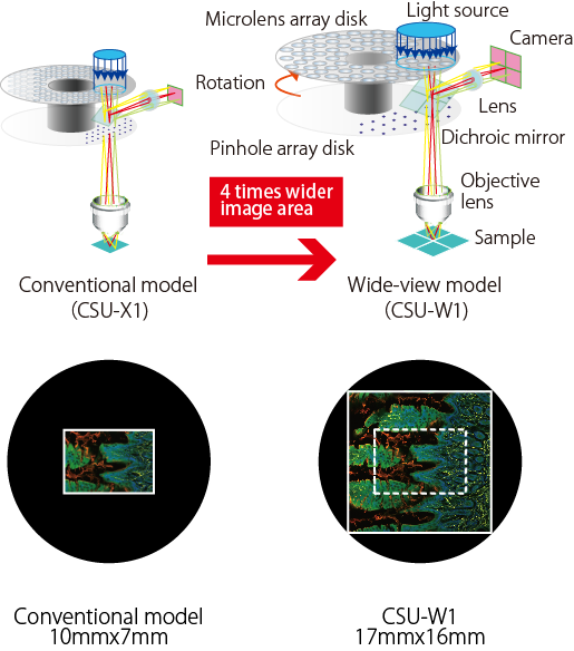
Image Credit: Yokogawa Corporation of America
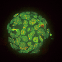
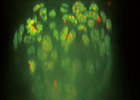
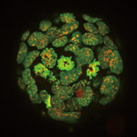
Clear
The image quality is significantly better with the newly designed disk unit. CSU-W1 makes it possible to see much deeper into thick samples because of the greatly decreased pinhole crosstalk.
Conventional model
XY MIP (left) and XZ Slice (right)
CSU-W1
XY MIP (left) and XZ Slice (right)
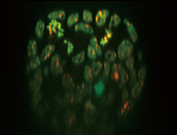
Mouse ES cell colony;Fluorescent probe: H2B-EGFP(Excitation:488 nm)
mCherry-MBD-NLS:(Excitation: 561 nm)
Objective lens: 60× silicone;
Z-sections/stack: 100 µm(0.4 m/251 slices)
Image Credit: Jun Ueda, Ph.D. and Kazuo Yamagata, Ph.D., Center for Genetic Analysis of Biological Responses, The Research Institute for Microbial Diseases, Osaka University
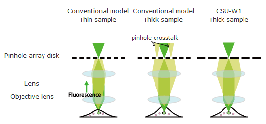
Image Credit: Yokogawa Corporation of America
Flexible
Functions that can be chosen with flexibility to suit a variety of applications.
High confocality pinhole (optional component)
The Company offers a 25 m pinhole size with higher confocality in addition to the standard 50 µm pinhole size.
Users can choose between the two pinhole sizes with an easy-to-use motorized disk exchange mechanism.
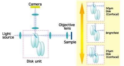
Image Credit: Yokogawa Corporation of America
New bright field through path (standard)
Phase contrast and other confocal and non-confocal images can be projected much more easily, thanks to a new mechanism that moves the disks out of the light path.
Simultaneous dual-color imaging mechanisms (T2 and T3 models)
In addition to the dual-camera models, which are significantly better than those for the CSU-X1, the CSU-W1 also offers a single-camera split-view model. Even the split-view provides twice the image area of the older model because of the wide field of view.
The split-view and two-camera models employ different dichroic mirrors, allowing for the selection of different dye combinations for dual-color imaging.
Details
In addition to two pinhole sizes, options for near-infrared observation, and an external light path that is helpful for various applications like photobleaching, CSU-W1 offers three basic configurations in total. A bright field light path is now a standard feature. The CSU-W1's switching mechanisms are all fully motorized, making them suitable for automated testing.
Basic configurations
CSU-W1 offers three basic multi-color imaging configurations: 1) split-view two-color imaging with one camera shared by two optical paths; 2) simultaneous two-color imaging with two cameras; and 3) sequential imaging with one camera and a filter wheel. Following installation, all features can be upgraded.
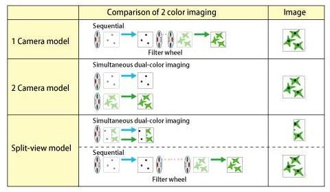
Image Credit: Yokogawa Corporation of America
Filter
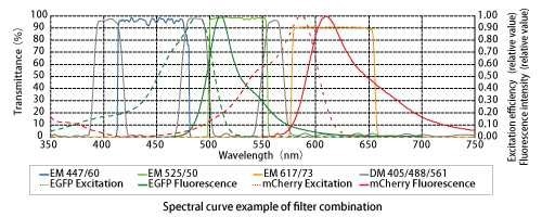
Image Credit: Yokogawa Corporation of America
Option
SoRa disk
A super-resolution method based on spinning-disk confocal technology has increased optical resolution by about 1.4×. Moreover, deconvolution is used to achieve a final resolution roughly twice that of the optical limit.
Uniformizer
The best choice for uniform CSU-W1 illumination. Uniform and effective lighting over the whole large field of vision.
Near Infrared (NIR) port
The NIR port offers excitation capabilities up to 785 nm to enable less invasive deep imaging. The NIR laser is introduced through a specialized optical fiber like visible lasers. The CSU-W1 unit can integrate visible and NIR lasers to enable simultaneous excitation.
External light path
An external light path provides a direct route to the microscope, avoiding the disks. This port allows for the introduction of an external light scanner for various applications, including photoactivation.
Lens switcher
A recently developed motorized lens switcher between two relay lenses helps adjust the CSU-W1 image size with different kinds of cameras and rapidly changes the magnification without changing the objective lenses.
Variable aperture
The CSU-W1's variable aperture helps reduce laser damage to the specimen by altering the laser illumination area and, consequently, the imaging area.
Selectable option
Source: Yokogawa Corporation of America
| Option |
1 Camera model |
2 Camera model |
Split-view model |
| NIR port |
× |
| External light path |
× |
| Variable aperture |
× |
N/A |
| Camera port lens |
Selectable from
0.83x, 1x |
Selectable from
0.83x, 1x
(1 Camera)
Selectable from
0.83x, 1x
(2 Camera) |
Selectable from
0.83x, 1x |
Additional lens
to lens switcher |
Selectable from 0.83x, 1x, 2x |
N/A |
2 Camera model, 1 Camera model
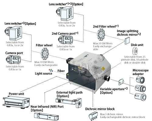
Image Credit: Yokogawa Corporation of America
Split-view model
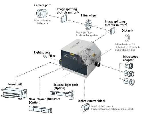
Image Credit: Yokogawa Corporation of America
*1 2 Camera model
*2 2 Camera model and Split-view model
*3 1 Camera model and 2 Camera model
*4 Under development
External dimensions
Image Credit: Yokogawa Corporation of America
Microscope-setup
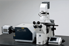
Zeiss Axio Observer. Image Credit: Yokogawa Corporation of America
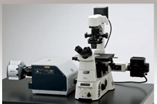
Nikon ECLIPSE Ti. Image Credit: Yokogawa Corporation of America
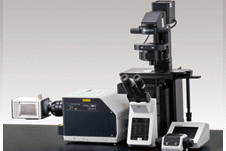
Olympus IX83. Image Credit: Yokogawa Corporation of America
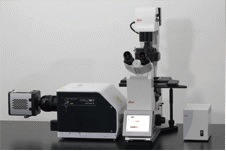
Leica DMi8. Image Credit: Yokogawa Corporation of America
General specifications
Source: Yokogawa Corporation of America
| General Specifications |
| Model |
1 camera model (T1) |
2 camera model (T2) |
Split-view model(T3) |
| Confocal scanning method |
Microlens-enhanced Nipkow disk scanning |
| Spinning speed |
1500 rpm - 4000 rpm Max 200 fps |
| External synchronization |
Scan-speed synchronization through pulse signals Input/output: TTL level 300 Hz up to 800 Hz |
| Disk unit |
Selectable up to 2 disks from 50 um
(for high magnification)
and 25 um (for low magnification)
Motorized switching |
| Bright field |
Motorized exchange between confocal and brightfield |
| Effective FOV |
17×16 mm |
| Excitation wavelength |
405 nm-785 nm |
| Laser introduction |
Yokogawa's standard fiber*1 VIS Laser port (405-647 nm)
【Option】NIR Laser port (685-785 nm) |
| Observation wavelength |
420 nm-850 nm |
| Dichroic mirror switching |
Motorized 3CH (Dichroic mirror block can be exchanged) |
| Emission filter wheel |
10-position filter wheel
Filter size φ25 mm
Switching speed*2: 100 msec max. (Standard mode) 40 msec max. (High-speed mode) |
6-position filter wheel
Filter size φ25 mm
Switching speed*2: 100 msec max. |
| External control |
RS-232C (CSU-X1 command upper compatible) |
| Microscope mount |
Yokogawa original |
| Camera adaptor |
C mount 1x (Variable magnification:0.83x ) |
| Light introduction port |
【Option】Photo breach etc |
| Operating environment |
15-30 oC、20-75% RH No condensation |
| Power consumption |
Input: 100-240 VAC ±10% 50-60 Hz 250 VAmax |
| External dimension |
Main unit |
327.1 (W) x
251.5 (D) x
475.1 (H) mm |
471.6 (W) x
251.5 (D) x
475.1 (H) mm |
420.8 (W) x
251.5 (D) x
373.6 (H) mm |
| Power unit |
225.4 (W) x 151.9 (D) x 378.3 (H) mm |
| Weight |
Main unit |
17 kg |
20.5 kg |
18 kg |
| Power unit |
4.5 kg |
| Attachable microscope |
Olympus IX series, Nikon ECLIPSE Ti, Zeiss Axio Observer, Leica DMI series*3 |
*1 Each CSU-W1 head is optimized with its fiber at factory. Please inquire about fiber exchange if necessary.
*2 Adjacent position.
*3 Some microscopes/options could limit the FOV of CSU-W1 or connection with CSU-W1, please inquire.