The Yokogawa CSU-W1 SoRa Confocal Scanner Unit is perfect for super-resolution live cell imaging, enabling researchers to capture high-speed, real-time images without the need for extensive sample preparation. Its spinning disk technology boosts the optical limit by approximately 1.4×, nearly doubling the resolution potential. Additionally, the newly released CSU-W1 Uniformizer further enhances imaging quality, providing even greater flexibility and performance for advanced research.
-
XY resolution surpasses the diffraction limit, reaching up to 120 nm
-
Ideal for super-resolution live cell imaging
-
User-friendly technology that’s easy to operate
-
Fully upgradable from the CSU-W1 for enhanced capabilities
XY resolution of approximately 120 nm
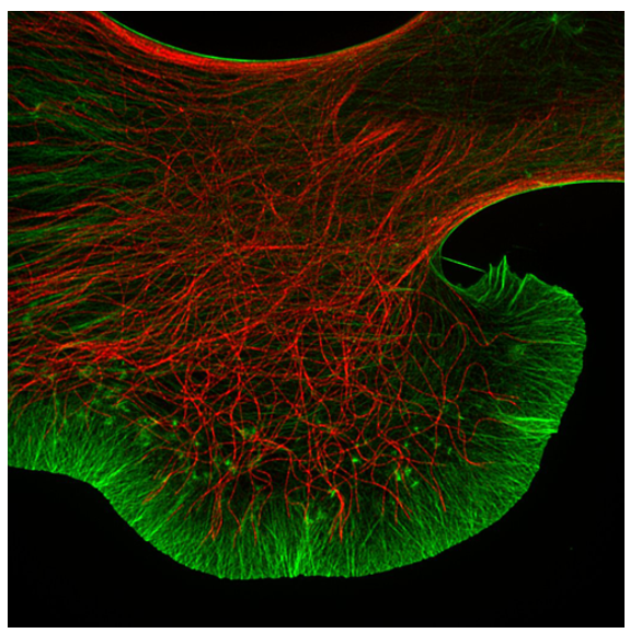
NG108 cell. Image Credit: Dr. Kaoru Kato, Biomedical Research Institute, National Institute of Advanced Industrial Science and Technology (AIST)
Spining-disk confocal technology has enhanced XY resolution by around 1.4 times the optical limit.
Moreover, deconvolution is used to achieve a final resolution roughly twice the optical limit.
Super-resolution live cell imaging system
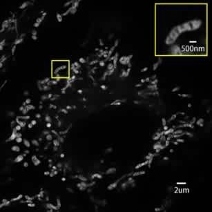
Real-time live cell imaging of mitochondria (10FPS). Image Credit: Dr. Kaoru Kato, Biomedical Research Institute, National Institute of Advanced Industrial Science and Technology (AIST)
Enable advanced live-cell imaging using standard fluorophores—no special dyes or fixation required.
Powered by the W1 Microlens Dual Disk Design, the system reduces phototoxicity and photobleaching while maintaining high imaging speed and resolution.
The CSU is easy to use
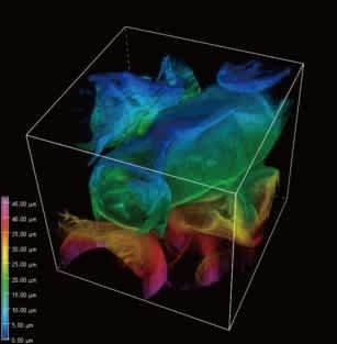
Image Credit: Yokogawa Corporation of America
Real-time viewing of high-resolution images is possible without the need for sample preparation.
Deep position observation is made feasible by optical sectioning using confocal technology.
Details
SoRa super-resolution principle
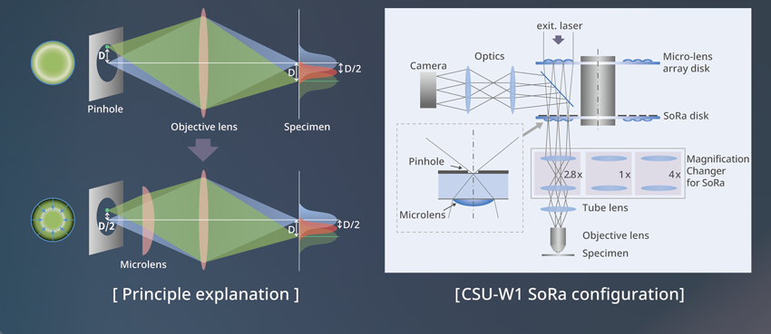
Image Credit: Yokogawa Corporation of America
In conventional confocal microscopes, image formation is represented as the product of illumination PSF (point spread function) and detection PSF. When analyzing the image created on the pinhole at position D from the optical axis, it is the product of the illumination and detection PSFs (as illustrated), and it is easy to see that information is conveyed at position D/2 from the optical axis at the light source.
Information from the D/2 position at the light source is magnified to D on the pinhole. To remedy this, a microlens is used, and the individual focal points projected onto the pinhole are optically reduced by half, resulting in an ideal image formation.
By doing so, the resolution is similar to that of an ideal confocal microscope, with the pinhole lowered to an infinitesimal size, resulting in an estimated 1.4× improvement over standard confocal microscopes.
Upgrading from the CSU-W1
Configuration when upgrading from the CSU-W1
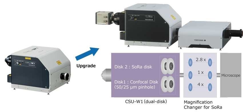
Image Credit: Yokogawa Corporation of America
The CSU-W1 can be equipped with a SoRa disk.
Users can perform imaging customized to their experimental needs by alternating between standard confocal observation and super-resolution observation with a SoRa magnification changer.
Source: Yokogawa Corporation of America
| . |
. |
| 1x: |
Confocal observation (CSU-W1) |
| 2.8x: |
Super-resolution 100x objective lens |
| 4x: |
Super-resolution 60x objective lens |
Product specification
Source: Yokogawa Corporation of America
| Product specification*1 |
| Model |
1 camera model (T1) |
2 camera model (T2) |
Split view model (T3) |
| Loadable model |
A SoRa disk can be loaded as disk 2, and disk 1 can be selected (50 μm or 25 μm) |
| Excitation wavelength |
405 nm~640 nm |
| Observation wavelength |
420 nm~680 nm |
| Effective scan field of view |
Depends on the magnification changer for SoRa specification (see below) |
| External light / NIR port |
An external light port cannot be equipped at the same time as the intermediate magnification switcher
The NIR port cannot be used together with a SoRa disk |
| Magnification changer for SoRa specification |
| Lens-switched light path |
3 light paths switched electronically 1x, 2.8x, 4.0x magnification |
| External dimensions |
425 (W) × 301.1 (L) × 122.5 (H) mm (excl. protrusions and supporting column) |
| Weight |
13 kg |
| Microscope connection |
Manufacturer-specific adapter |
| Resolution: : PSF FWHM*2 |
| XY/Z resolution (optical super-resolution) |
150 nm / 320 nm |
|
| XY/Z resolution (after deconvolution) |
120 nm / 300 nm |
*1 Only items which differ from the CSU-W1 are shown. Specification : Confocal scanner unit CSU-W1
*2 Resolution value is for reference only.