The Single Cellome Unit SU10 allows materials, like genome editing tools, to be delivered directly to the cytoplasm or nucleus of specific single cells. It is also capable of single-cell sampling. The SU10 is for use with stereo microscopes and inverted optical microscopes; it does not come with a microscope.
Advantages of SU10
- Single-cell delivery that enters the cytoplasm or nucleus directly
- Nano-point sampling from the cytoplasm or nucleus
- Cell damage is minimal
- High speed, high success rate, and automated
- Injection through cell wall of plants for gene editing
Feature
Single-cell targeting with direct delivery into the nucleus or cytoplasm
- Deliver materials into the targeted cells' cytoplasm or nucleus while selecting cells for delivery under a microscope.
- The software allows for simple XY-position control and automatic Z-position delivery control.
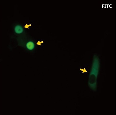
FITC-labeled dextran solution (molecular weight 70,000) was delivered into Human iPS cells (201B7, 2D culture). Image Credit: Matsuzaki Lab, RIKEN
Minimal damage to cells
- The nanopipette is a glass pipette that minimizes cell damage by having a minimum tip outer diameter of a few tens of nanometers.
- Live single-cell analysis is made possible by delivery with high cell viability.
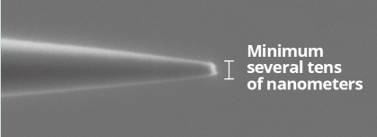
The tip of the nanopipette under an electron microscope. Image Credit: Yokogawa Corporation of America
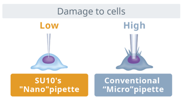
Image Credit: Yokogawa Corporation of America
Automated, high speed, and high success rate
- The SU10 delivers, inserts, and detects cell surfaces automatically. With a 90% success rate, the process takes about 10 seconds.
- With SU10, tasks previously completed by skilled researchers by hand are considerably easier to handle.
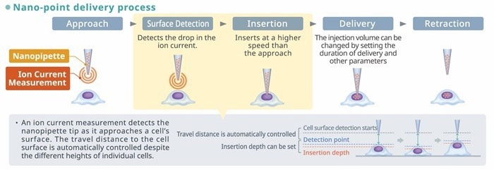
Image Credit: Yokogawa Corporation of America
Nano-point sampling
- It is possible to take a tiny sample from a particular cell area.
- Applications like genetic analysis can make use of collected samples.
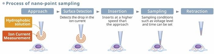
Image Credit: Yokogawa Corporation of America
SU10 automated nano-point delivery operation procedures
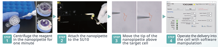
Image Credit: Yokogawa Corporation of America
Application example
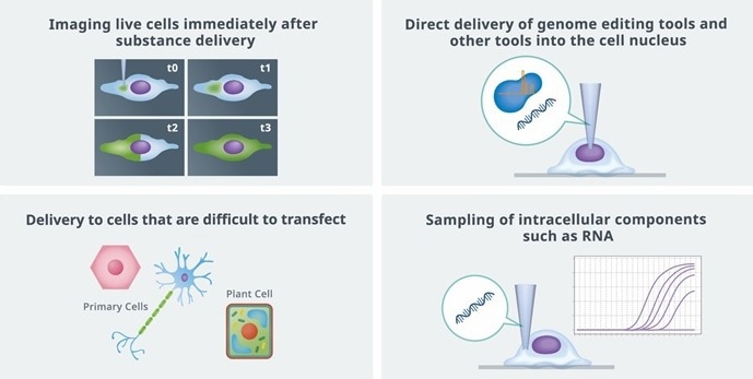
Image Credit: Yokogawa Corporation of America
Application example: Delivery of genome editing tools
Using SU10, Cas9RNP and Cas9RNP+donor DNA (100b) were delivered to HeLa cells that expressed GFP, and the GFP gene was successfully knocked out or modified to the BFP gene.
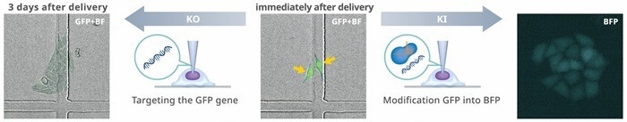
Image Credit: Kamakura Laboratory, Department of Applied Biological Science, Faculty of Science and Technology, Tokyo University of Science
Application example: Delivery of primary cells
Greatly increases transfection effectiveness in cells that have proven challenging to transfect.
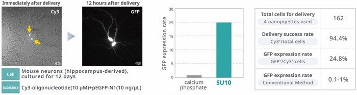
Image Credit: Ms. Kanako Iwasaki, Yasushi Okada Lab, The University of Tokyo
Application example: Delivery to cultured plant cells and plant tissues
- Depending on the sample, the SU10 can be used with a stereo microscope for thick tissues and an inverted microscope for cultured cells.
- Suitable for cells with high turgor pressure or stiff cell walls
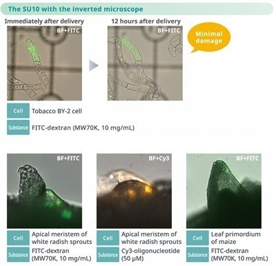
Image Credit: Dr. Daisuke Kurihara, Higashiyama Group, The Institute of Transformative Bio-Molecules, Nagoya University
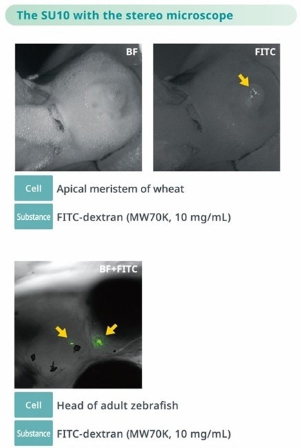
Image Credit: Dr. Koji Ando and Dr. Kazu Kikuchi, Department of Cardiac Regeneration Biology, National Cerebral and Cardiovascular Center
Verified SU10 performance applications
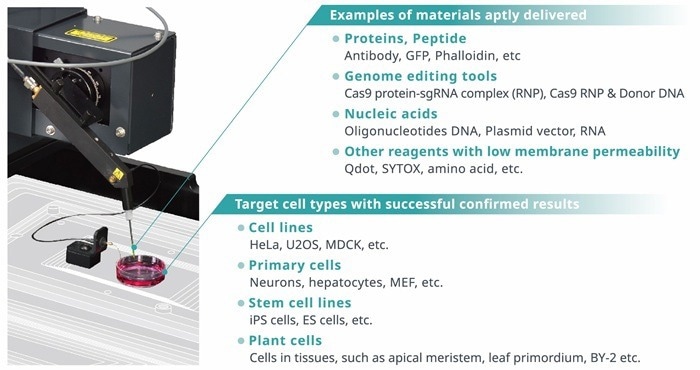
Image Credit: Yokogawa Corporation of America
Injection movies
Available to separate the nucleus and cytoplasm
Cell: HeLa
Substance: FITC-dextran
Image Credit: Yokogawa Corporation of America
Live cell imaging of the movement of delivery reagents from the cytoplasm to the nucleus
Cell: HeLa
Substance: SYTOX® Green, 500 μM
Image Credit: Yokogawa Corporation of America
Enables antibody staining of intracellular molecules/organelles in live cells
Cell: U2OS
Substance: Alexa647-conjugated anti-TOMM20 antibody
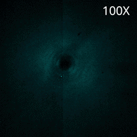
Image Credit: Yokogawa Corporation of America
Imaging of intracellular environments
Cell: HeLa
Substance: Qtracker 655, 1 µM
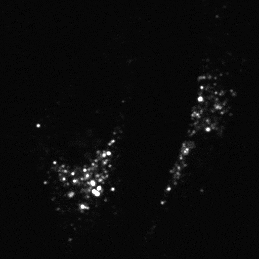
Image Credit: Yokogawa Corporation of America
Non-membrane permeable reagents can be injected, enabling live cell imaging of actin
Cell: Fish epithelial keratocyte
Substance: Alexa Flour 488 Phalloidin
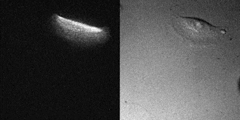
Image Credit: Yokogawa Corporation of America
Operation principle
The technology is based on SICM and involves automatic cell detection and penetration.
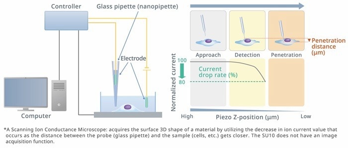
Image Credit: Yokogawa Corporation of America
Electro-osmosis and electrophoresis are methods used to deliver substances and solutions into cells.
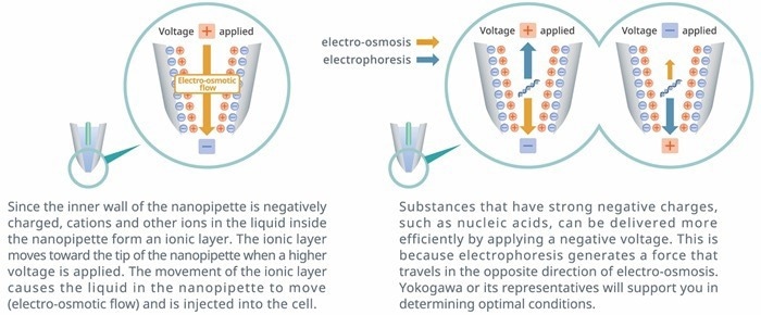
Image Credit: Yokogawa Corporation of America
Specification
Source: Yokogawa Corporation of America
| . |
. |
. |
Motor
actuator |
Number of axes |
XYZ direction: three axes |
| Stroke |
XZ: Max. approximately 50 mm, Y: Max. approximately 30 mm |
| Setting resolution |
0.625 um |
| Maximum movement speed |
1 mm/sec |
Piezo
actuator |
Number of axes |
Z direction: one axis |
| Stroke |
500 um |
| Setting resolution |
10 nm |
Retraction
stage |
Number of axes |
X direction: one axis |
| Stroke |
100 mm |
Rotary
stage |
Range of movement |
360° |
Joystick
Controller |
Number of joysticks |
For XY operation: one piece, For Z operation: one piece |
| Number of switches |
2-selection toggle switch: one piece, 3-selection toggle switch: one piece |
Power
voltage |
Power voltage |
100 to 240 V AC |
| Power frequency |
50/60 Hz |
| Power consumption |
70 VA |
External
dimensions
and weight |
Main unit |
337 to 456 (W) mm × 230 (H) mm × 377 (D) mm, 7.4 kg |
| Main controller |
133 (W) mm × 309 (H) mm × 364 (D) mm, 5.9 kg |
| Joystick controller |
140 (W) mm × 114 (H) mm × 144 (D) mm, 1.2 kg |
| nanopipette |
Tip outer diameter: Several tens of nanometers or less (reference value) (in case of SU10ACC-NP02) |
Ambient
operating
conditions |
Temperature |
Main unit and electrode head: 5 to 40 °C
Other modules: 15 to 35 °C |
| Humidity |
Main unit and electrode head: 80% RH or less (without condensation)
Other modules: 20 to 70% RH (without condensation) |
| Installation environment |
Keep the system away from direct sunlight. Do not allow it to come in direct contact with water, oil, organic solvents, etc. Make sure there are no flammable, toxic, or corrosive gases. Also, do not use or store the system in locations where sand, dust, or particles have accumulated, where there are sources of strong electromagnetic noises, where fire is used, that are readily exposed to water, or where strong vibrations occur. |
| Installation posture |
Horizontal installation |
| Altitude |
2000m or less |
| Storage environment |
Temperature |
-10 to 50 °C |
| Humidity |
95% RH or less (without condensation) |
| Installation environment |
Keep the system away from direct sunlight. Do not allow it to come in direct contact with water, oil, organic solvents, etc. Make sure there are no flammable, toxic, or corrosive gases. Also, do not use or store the system in locations where sand, dust, or particles have accumulated, where there are sources of strong electromagnetic noises, where fire is used, that are readily exposed to water, or where strong vibrations occur. |
| Operation Environment |
For use with an inverted optical microscope. * Microscope is not included with the SU10.
Please contact Yokogawa to possibly install the SU10 on a different inverted microscope.
Installation examples; Evident IX83, Nikon Ti2, Zeiss Axio Observer |
Software specifications
Installation PC requirements |
OS |
Windows 10, Windows 11 |
| USB |
One (1) or more Type A2.0 USB ports |