Incucyte® CytoLight Rapid Dyes for live-cell labeling enable simple, fast and highly efficient labeling of living cells. These dyes are well suited for identifying the location of cells within a population and monitoring the dynamic interactions between cells in mixed cultures.
They are ideal for labeling cell types that are difficult to label using BacMam, lentivirus or other transfection-based labeling approaches (e.g. immune cells and primary cells). These dyes can be multiplexed with Incucyte® Annexin V, Caspase-3/7 or Cytotox reagents for simultaneous readouts of apoptosis or cytotoxicity.
Key Advantages
- Simple to use – harvest cells, add dye, incubate 20 minutes, plate cells and image
- Rapid, high efficiency cell labeling
- Non perturbing to cells at optimized dye concentrations
- Ideal for cell types difficult to label with BacMam or lentivirus (e.g. primary and immune cells)
- Dyes do not pass to adjacent cells – ideal for monitoring cells in co-cultures
The Incucyte® CytoLight Rapid Dyes for live-cell labeling freely pass through cell membranes and into cells, where they are transformed into a cell membrane-impermeant form. The dyes are transferred and diluted in daughter cells, but are not transferred to adjacent cells in a population. At optimized concentrations the CytoLight Rapid Dyes are non-perturbing to cells and fluorescence can be measured up to 48 hours post labeling.
CytoLight Rapid Dyes are Non-Perturbing and Enable High-Efficiency Cell Labeling
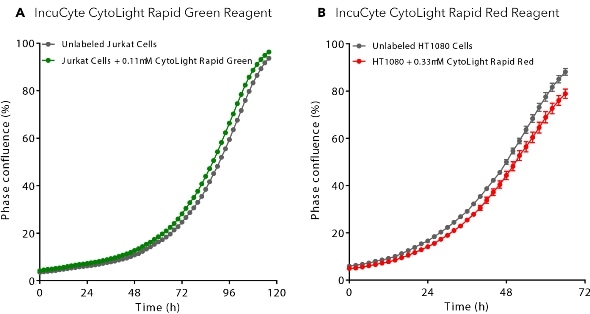
Optimized Dye Concentrations Do Not Perturb Cell Health: CytoLight Rapid Dye labeled Jurkat (non-adherent) and HT-1080 (adherent) cells show equivalent cell growth profiles to non-labelled cells.
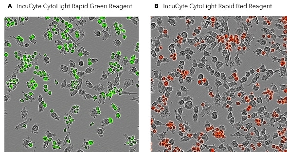
Rapid, High Efficiency Labeling of Adherent and Non-Adherent Cell Types: After a 20 minute incubation with the CytoLight Rapid Dyes, effectively all cells in the population (>95%) are fluorescently labeled. Note the homogeneous cytoplasmic labeling and the healthy cell morphology.
Visualize and Monitor Spatial and Temporal Cell Interactions
Detect the Dynamic Interactions Between Immune and Tumor Cells in Real Time: Peripheral blood mononuclear cells (PBMCs) were activated and labelled with Incucyte® CytoLight Rapid Green Dye and then added to tumor cells stably expressing a red fluorescent protein (CytoLight Red Lentivirus Reagent). Immune cells in close proximity to tumor cells are masked in yellow using Incucyte® image analysis software.
IncuCyte® CytoLight Rapid Green Dye
Monitor Viability in Mixed-Cultures and Multiplex with Cell Health Reagents
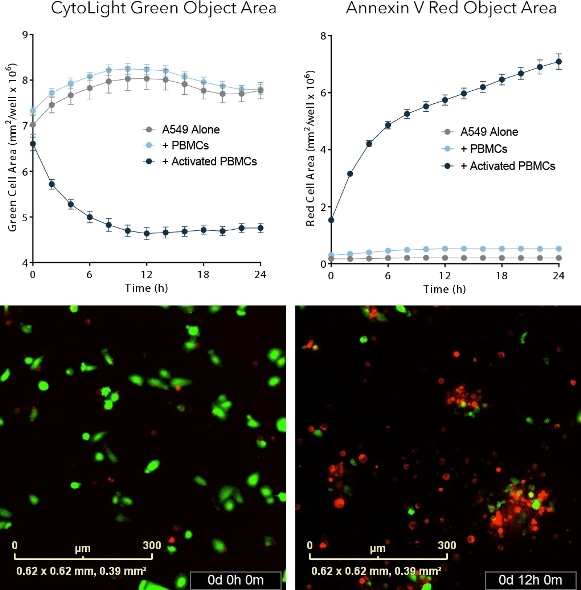
Analysis of Target Tumor Cell Viability Over 24 Hours Multiplexed with an Apoptosis Readout: Human adenocarcinomic alveolar basal epithelial cells (A549) were labeled with Incucyte® CytoLight Rapid Green Dye and cultured in the presence of pre-activated PBMCs and Incucyte® Annexin V Red reagent. Tumor cell killing by activated PBMCs is monitored in real time by measuring the loss of green object area (left) and increase of red object area (Annexin V signal) over time.
Monitor Cell Morphology and Compare Phenotype
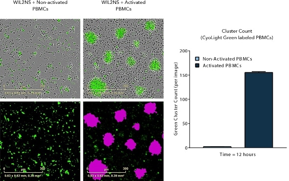
Visualize Changes in the Phenotype of Labeled Subpopulations: Incucyte® CytoLight Rapid Green labeled human PBMC’s in co-culture with WIL2-NS B lymphocyte cells. Rapid Green dye labeling enables visualisation and quantification of PBMC clustering when activated (anti-CD3/IL-2).