Bimolecular fluorescence complementation (BiFC) is a recent technique used in the investigation and direct visualization of protein–protein interactions (PPIs) and interaction between proteins and other macro-molecules that are essential for survival of cells. Identification of these relations specifies their working inside the cells in brief. Organisms with PPI networks include humans, worms, yeast, and plants.
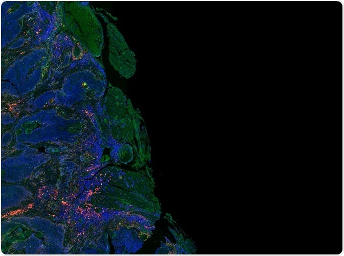
Credit: Carl Dupont/Shutterstock.com
BiFC is mainly used in genome-wide studies of PPIs; it is identified as an effective technique in uncovering new protein interactions, and providing novel information on functions of protein.
Principle of BiFC
The working principle of BiFC is based on the development of a fluorescent complex, as a result of the association of two segments of a fluorescent protein when they are in close proximity due to protein–protein interaction in the fragments, i.e., in BiFC, a fluorophore is divided into amino and carboxyl terminal ends. These ends are combined into two proteins. When these two proteins interact, both the segments re-associate resulting in re-formation of the fluorophore and fluorescence at the sites of interface.
Set of rules to design BiFC
The BiFC experiments – as it is based on the interaction between the fluorescent protein segments - takes place only under certain circumstances. BiFC analysis involves an assay that is fabricated in such a way that it satisfies all the parameters affecting the association of the fluorescent protein segments. The procedure of assay fabrication includes:
BiFC calibration
BiFC calibration is carried out in four stages as follows:
- Selection of appropriate fluorophore that works finely as BiFC synthesis partners. Examples of fluorophores are Venus and yellow fluorescent protein (YFP).
- Labeling of proteins in which the BiFC segments are bonded to the amino- or carboxyl-terminals of the selected protein.
- Determination of transfection circumstances before the testing of multiple mutants.
- Determination of changes that occur in the localization of proteins due to addition of BiFC segments.
Plating and cell transfection
In this process, the cells are initially plated in a glass bottom plate and in two wells of a 6-well plate and then they are allowed to settle overnight at room temperature. Then the cell DNAs are prepared for transfection process. After completion of the transfection process, the combination is divided equally and then incubated at room temperature for a few hours. Then the transfection media is removed and incubated once again at room condition.
Preparation of cells for imaging
In this process, the cells are initially analyzed using an epifluorescent microscope in order to check their fluorescence. Then, the preparation of cells is done by addition of paraformaldehyde after which the cells are lysed to obtain the image.
Imaging of cells
The intensity of fluorescence is determined for every single cell. Selective analysis of BiFC signals is achieved by performing transfection along with CFP. Imaging is done using a confocal microscope.
Fluorescence quantitation
Measurement of the intensity of fluorescence is accomplished using several imaging software.
Analysis of data
The efficiencies of fluorescence complementation are determined by the value obtained by division of the intensities of fluorescence complementation and the intensities of whole fluorescent protein in each cell.
Design of multicolor BiFC experiment
Multicolor BiFC experiment involves the combination of substitutional partners and the segments of fluorescent proteins that yield compounds with different color bands. Sequential imaging is achieved by visualization of the compounds involving distinct emission and excitation wavelengths. Imaging of the compounds is done alternatively to prevent re-localization of either compound based on the lag between the imaging of the compounds.
The fluorescence intensities should be maintained at constant magnitude in order to prevent development of variation in the distributions caused by variation in the signal-to-background ratio. The disturbances between two fluorophores can also be rectified by imaging the cells that represent only one group of proteins.
Multicolor BiFC was first established in mammalian cells in studies that used various combinations of YFP and CFP. BiFC contrives to examine the relative efficiencies of bFos interaction with bJun and bATF2.
The improvement of a multicolor BiFC method for plant cells helps in the investigation of simultaneous development of alternative calcium sensor/protein kinase complexes in planta.
Applications of the BiFC assay
Applications of BiFC include the imaging of steady-state distribution of compounds developed by grouping of proteins in various types of cells and organisms. It also enables the imaging of interactions between multiple proteins in a single cell in addition to alterations in the covalent proteins that provide additional information about the biological processes of protein compounds that are recently encountered.
It is also helpful in the identification of various protein segments that can be combined by the interaction between the proteins that are bonded to the segments that include β-galactosidase, ubiquitin, and dihydrofolate reductase. BiFC experiments are also helpful in the determination of topology of membrane proteins and high input and output inspection of small molecular effects on the protein compounds.
Further Reading
Last Updated: Jul 20, 2023