The seemingly effortless and instantaneous color and iridescence changes displayed by octopuses, squid, and cuttlefish are one of Nature’s most amazing sights. Now, researchers have discovered how to use the cephalopod proteins responsible for these changes to manipulate the optical refractive index (RI) of mammalian cellular compartments.
By changing this intrinsic physical property of the cell, microscopists could soon be using the RIs of live cells as molecular probes and capturing the resulting label-free quantitative phase images using Tomocube’s holotomography microscope.
Gerald M Pao(1) and his colleagues from the Salk Institute for Biological Studies in California set out to genetically manipulate the RI of living cells by synthesizing known cephalopod reflectin genes and cloning new reflectin genes expected to yield a higher RI from the wild Glitter or Bigfin reef squid (Sepioteuthis lessoniana).
Epitope tagged for antibody recognition with either an HA or FLAG-tag, with an added preprotrypsin signal sequence to target the protein into the secretory pathway, a C-terminal KDEL ER retention signal was added to specifically target the endoplasmic reticulum (ER) of cells. Their success is demonstrated in the quantitative phase-contrast microscopy studies, where the Tomocube holotomography microscope measured an RI of up to 1.4, well above the ER RI of 1.35.
Microscopists will tell you that RI is an intrinsic physical property of any material, including cellular components, and is not generally considered amenable to manipulation. This fantastic work from Gerald Pao and his Salk colleagues ends this notion and opens up a new era of control of the RIs of live cells. In essence, they have shown how to turn the cell itself into a molecular probe for quantitative phase imaging.
One use of this new technology may be to manipulate the RI to achieve transparency of scattering tissues through refractive index matching. Until now, this has required fixed, permeabilized whole organ samples, which precludes live imaging. However, with a genetically encoded protein, this may be conceptually possible.”
Aubrey Lambert, Tomocube’s Chief Marketing Officer
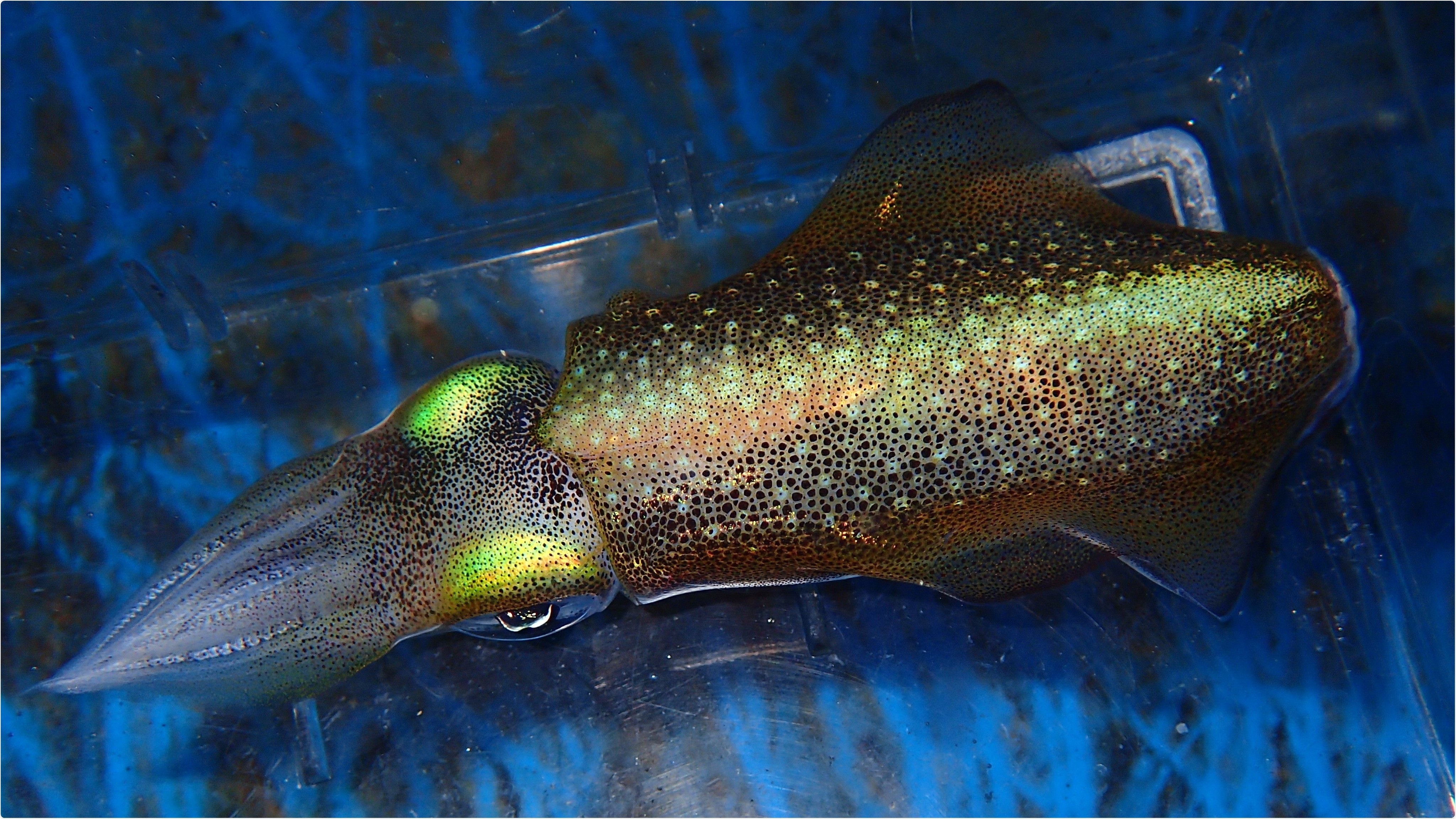
Reflectins proteins are the main components responsible for the structural coloration and iridescence in cephalopods, mainly used for camouflage. They are found in specialized cells, the iridophores, forming Bragg mirrors by alternating layers of high and low refractive index material.
Within the membrane stacks, reflectins form nanoparticles of < 5nm to >500nm and confer the high optical refractive index property to iridophores. Reflectins are also utilized by cephalopods for white light scattering and to achieve chromatic invariance of chromatophores, pigment sacs, as these expand or contract.
The Tomocube holotomography microscope delivers quantitative, nanoscale, real-time, label-free 3-D images of individual living cells quickly and simply without any sample preparation. The holotomography images also deliver vital information on unique cell properties, including cell volume, shapes of sub-cellular organelles, cytoplasmic density, surface area, and deformability.
The latest HT-2 model combines the quantitative phase imaging (QPI) approach of label-free, 3-D refractive index (RI) tomography with 3-D fluorescence imaging.
Winner of the Microscopy Today 2019 Innovation Award, this microscope enables long-term tracking of specific targets in live cells while minimising stress. The capability to easily deliver holotomography and fluorescence correlative analysis in 2D, 3D and 4D will enable researchers and clinicians to open new frontiers in bioscience and better understand, diagnose, and treat disease.
 Tomocube releases first of it's kind, 3D fluorescent holotomography microscope