News Medical-Life Sciences sat down with Dr Mhairi Morris, Reader in Exercise Oncology at Loughborough University, working within the School of Sport, Exercise and Health Sciences, to discuss the ways that exercise can impact cancer cell behavior.
Image Credit: Promega Corporation
Can you introduce yourself and your role at Loughborough University within the School of Sport, Exercise and Health Sciences?
I'm a Reader in Exercise Oncology at Loughborough University, working within the School of Sport, Exercise and Health Sciences. The school covers a broad range, from exercise physiology through to molecular biosciences. My own research sits at the intersection of exercise science and oncology, with a specific focus on breast cancer.
What is the focus of your research on exercise and breast cancer, and why is this area important to study?
My research focuses on understanding how exercise affects cancer cell behavior within the breast cancer microenvironment, specifically in the context of obesity. My team is investigating whether exercise can counteract the negative influence that fat cells have on cancer cell viability.
The importance lies in understanding whether exercise could serve as a therapeutic tool to mitigate some of the pro-tumorigenic effects of adipose tissue on cancer progression.
What led your team to investigate the role of adipose tissue in the tumor microenvironment?
I saw Elvira Weber (Head of the laboratory at CURE 3D) speak at WORD+2024 and was fascinated by the idea of "adipoids" (adipose spheroids), which led me to explore the field of cancer-associated adipocytes. The idea that exercise might mitigate their effects in the breast tumor microenvironment intrigued me.
My research is premised on the known relationship between obesity, adipose tissue, and cancer outcomes. We are exploring how fat cells from two distinct sources interact with cancer cells, reflecting broader scientific interest in the obesogenic tumor microenvironment.
Can you describe the 3D model you have developed to study the obesogenic tumor microenvironment?
We have developed a 3D model of the obesogenic breast cancer microenvironment that incorporates fat cells from two different sources: visceral fat, the "bad fat" found around the abdomen, and subcutaneous fat, which I think of as the "good fat" that burns energy.
Cancer cells are cultured within this 3D system alongside these adipocytes and embedded in an extracellular matrix, replicating the interactions that would occur in the natural tumor microenvironment in vivo.
Why was it important for your research to move from traditional 2D systems to a 3D culture model?
3D models are far more representative of what happens in vivo than 2D models. I like to use a football analogy: 2D cell culture is like playing football lying flat on the ground side by side, unable to move (technically possible, but wholly unrepresentative of a real match). In 2D, cells can only interact side by side, whereas in 3D, they interact across all three dimensions.
Crucially, 3D models also allow other cell types and extracellular matrix components to be incorporated, enabling a more authentic recreation of the tumor microenvironment.
What challenges do complex, matrix-embedded 3D systems present when measuring cell viability?
Traditional assays such as MTT and WST-1 required multiple additional steps to digest the matrix before the assay could even be run. When we moved from 2D to 3D systems, we also found we could not reliably replicate results from batch to batch or experiment to experiment, a significant reproducibility problem that really undermined our confidence in the data.
What led you to adopt the CellTiter-Glo® 3D Cell Viability Assay for this application?
After experiencing poor reproducibility with the MTT and WST-1 assays in 3D systems, I was introduced to Promega’s CellTiter-Glo 3D: it was a revelation. It delivered consistently reproducible results across experiments and batches in a way that my previous assays never had, and that reliability was the primary reason for making the switch.
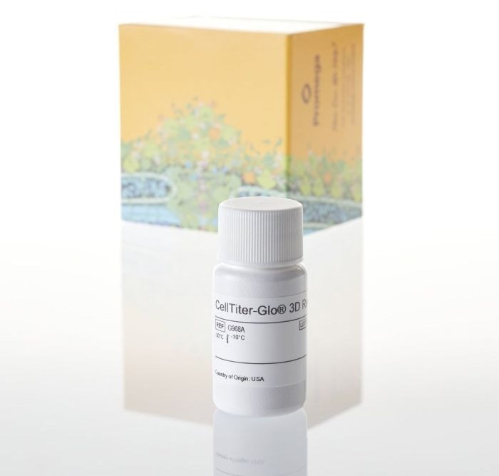
Image Credit: Promega Corporation
How does the CellTiter-Glo® 3D assay integrate into your workflow when working with 3D cultures?
It is very straightforward to integrate. CellTiter-Glo 3D is a lytic reagent, so it is added directly to the wells where it lyses cells and makes them release ATP. That ATP then drives the luciferin-luciferase reaction, producing a luminescent signal proportionate to the number of metabolically active, viable cells.
In practice, we add the reagent to each well, place it on a plate shaker for five minutes, incubate in the dark for 25 minutes, and then read the results.
What advantages have you observed in terms of reproducibility, sensitivity, or ease of use with this assay?
There are three key advantages I would highlight. First, reproducibility: I now consistently achieve comparable results across batches and experiments, which I simply was not able to do with previous assays. Second, ease of use: adoption required minimal optimization beyond standard project-specific adjustments.
Third, time efficiency: compared to the four-hour incubation required by the MTT assay, CellTiter-Glo 3D only takes around 30 minutes from start to result, which significantly accelerates throughput. If I had to summarize the assay in three words, I would say: easy, reliable, and inexpensive.
How well does the assay perform when working with matrix-embedded or co-culture systems?
This is honestly one of the most practically valuable features. Unlike other assays that require several digestion steps to break down the matrix before you can proceed, CellTiter-Glo 3D can be applied directly to matrix-embedded spheroids and organoids without any prior digestion. That was an unexpected but very welcome benefit.
In addition to viability, how are you assessing cancer cell proliferation in your model?
More recently, I have begun using the Lumit hKi-67 immunoassay to assess cell proliferation alongside our viability measurements.
What key findings have you observed regarding the impact of adipocytes and exercise on cancer cell viability and behavior?
When cancer cells are co-cultured with fat cells, we observe enhanced cancer cell viability, suggesting that adipocytes promote tumor cell survival. However, when we overlay exercise on that system, through exercise-conditioned media, we see a reduction in viability. The early finding is that exercise may be able to negate some of the pro-tumorigenic effects that fat cells exert on cancer cells within the tumor microenvironment.
What are the next steps in your research, particularly in understanding the mechanisms behind exercise-driven changes in the tumor microenvironment?
Now that the model has been established, our next priority is to use it to begin unpicking the mechanistic details underlying the effects we have observed with exercise and cancer, moving from simply observing that exercise reduces viability to understanding precisely how and why it does so at the molecular and cellular levels. It is a genuinely exciting phase of the work.
About Dr Mhairi Morris
Mhairi A. Morris, PhD, is a Reader in Exercise Oncology at Loughborough University, UK, where she teaches cancer biology and conducts research into the obesogenic breast cancer microenvironment using novel 3D co-culture models to explore the effects of exercise and endocrine-disrupting chemicals on breast cancer growth and metastasis. Mhairi earned a PhD in tumour virology at the University of Birmingham in 2009 and pivoted her research focus to exercise oncology in 2017 after joining Loughborough University. Outside of academia, Mhairi is a certified Les Mills Dance™ and BodyJam™ instructor, combining her enthusiasm for exercise with her academic pursuits.
About Promega Corporation
At Promega Corporation, creativity and connection drive discovery and innovation. We serve our customers by providing over 4,000 solutions for genomic, protein, cellular analysis, drug discovery and genetic identity research.
From large-scale production to meeting specific quality or regulatory requirements to creating custom formulations, our custom and OEM customers benefit from the priority we place on authentic interactions with our customers, partners, and vendors. We build connections across the globe and in our local communities because creative problem solving requires a network of diverse ideas and viewpoints.
Our tailored R&D solutions provide customers with expert support from start to finish for small molecule and biologics drug discovery and development workflows. We handle over 85% of our manufacturing, which enables superior quality control, more customization options, and enhanced customer support.
Beyond our products, Promega stands out as a thought leader, embracing sustainable practices and supporting our global community. We commit to ensuring that our solutions not only meet the highest standards of quality and integrity but also remain adaptable to the ever-evolving landscape of scientific research.
Discover more about how Promega is making science easier, more reproducible, and more powerful at Promega.com.
 Promega launches XpressAmp™ Direct Amplification Reagents