In this interview, AZoLife Sciences speaks with Boyd Butler, a microscopy and high-content screening expert at Molecular Devices, about the growing role of AI in high-content screening (HCS). Drawing on his experience as a scientist, core facility director, and microscopist, Butler discusses the importance of AI-ready image data, the challenges of imaging complex 3D models, and how advances in optics, automation, and machine learning are transforming drug discovery. He explains how AI-powered image analysis helps researchers generate more reliable results, identify subtle phenotypic changes, and scale imaging workflows, while highlighting the growing use of human-relevant 3D models and the importance of high-quality imaging data for effective AI applications.
Can you please introduce yourself and your role at Molecular Devices?
My background is in biophysics and optical physics, and I spent many years in academia as both a faculty member and core facility director. Throughout my career, microscopy has been a central focus, particularly in understanding complex biological systems and helping researchers extract meaningful information from imaging data.
Today, I work closely with high-content screening technologies and imaging workflows at Molecular Devices. My role involves helping scientists adopt advanced imaging approaches, including AI-assisted analysis, automation, and high-content screening platforms, supporting applications ranging from basic research to drug discovery.
Why does high-content screening need AI-ready image data?
AI systems in high-content imaging are only as good as the data you train them on. In other words, input quality directly determines output quality. This fundamental truth remains constant across domains: high-quality imagery leads to more accurate and meaningful AI results. By emphasizing data excellence from the start, we set AI up for success.
One of the most important applications of AI in high-content screening is segmentation. Before a model can classify or analyze a cell, it must first accurately identify the cell and delineate its boundaries, nucleus, Golgi apparatus, and other structures. To do that reliably, the imaging data must be of high quality.
Good optics and well-prepared samples are equally important. Even the most advanced imaging platform cannot compensate for poor sample preparation. High signal-to-noise ratios, clear cellular features, and robust metadata provide the foundation that AI models need to generate meaningful results.
This becomes particularly important in applications such as toxicity testing. Researchers are often looking for subtle phenotypic changes that indicate whether a compound may be harmful. If the model cannot accurately distinguish biological structures because of poor image quality, the entire analytical workflow suffers. High-quality, AI-ready image data is therefore essential for reliable segmentation, classification, and downstream biological interpretation.
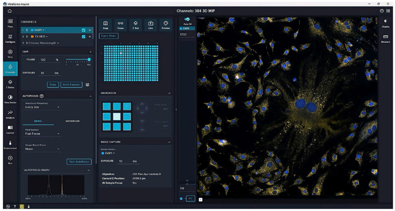
MetaXpress® High-Content Image Acquisition and Analysis Software offers an intuitive, user-friendly environment that is quick for novices to master while remaining flexible and customizable for advanced users. New features include easily controlled transmitted light acquisitions and identification, and centering “rare” objects in a well for high magnification acquisition. Image Credit: Molecular Devices.
What makes 3D models difficult to image and analyze at scale?
The future of biological research lies in human-relevant 3D models, including organoids and other advanced cellular systems. While traditional 2D cultures have been incredibly valuable, they do not fully capture the complexity of human biology.
The challenge is that 3D systems are significantly harder to image. Brightfield imaging struggles to penetrate thick samples, while even advanced imaging techniques face challenges related to heterogeneity, refractive index mismatches, and light scattering throughout the sample.
Organoids also generate enormous amounts of data. Instead of acquiring a single image plane, researchers may need to capture hundreds of optical sections throughout a 3D structure. When multiplied across entire microplates, data volume becomes a major challenge.
Analysis presents another layer of complexity. Segmenting cells in a flat monolayer is difficult enough, but segmenting structures within dense 3D tissue requires significantly greater image quality and computational power. Despite these challenges, 3D systems provide a much more physiologically relevant model for studying toxicity, drug penetration, and disease biology, making the effort worthwhile.
How do better optics improve high-content imaging results?
Better optics improve every stage of the imaging workflow, particularly when working with 3D samples.
High numerical aperture objectives provide improved resolution and enable researchers to distinguish structures that would otherwise appear merged together. Water immersion objectives are especially valuable because they reduce refractive index mismatches between the optical system and aqueous biological samples, resulting in clearer images and better signal quality.
Confocal imaging further improves results by eliminating out-of-focus light. This becomes particularly important in thick 3D samples, where background fluorescence can interfere with accurate image interpretation.
From an AI perspective, image quality directly influences model performance. If two closely positioned cellular structures cannot be resolved, segmentation models may incorrectly identify them as a single object. Poor resolution, therefore, affects not only visualization but also AI training, classification accuracy, and biological interpretation. High-quality optics provide the detailed information needed for robust segmentation and more reliable downstream analysis.
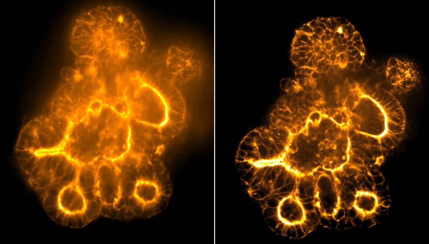
Image on left acquired with 60 µm pinhole confocal disk and image on right acquired with 50 µm pinhole deep tissue disk on the ImageXpress® HCS.ai High‑Content Screening System. Image courtesy of Molecular Devices.
How does automation increase throughput and reproducibility?
Automation improves speed and consistency.
In laboratory environments, automation can handle repetitive tasks such as moving plates between instruments, allowing researchers to collect data continuously with minimal intervention. This increases throughput while reducing the risk of human error.
Equally important is workflow automation. Standardized protocols ensure that researchers across different sites and laboratories follow the same procedures. This creates consistency within experiments and across organizations, which is especially important in pharmaceutical development, where reproducibility is critical.
Automation also reduces variability introduced by manual processes. Whether researchers are collecting images, setting acquisition parameters, or analyzing results, automated workflows help ensure that experiments are performed consistently every time. This accelerates discovery while increasing confidence in the data being generated.
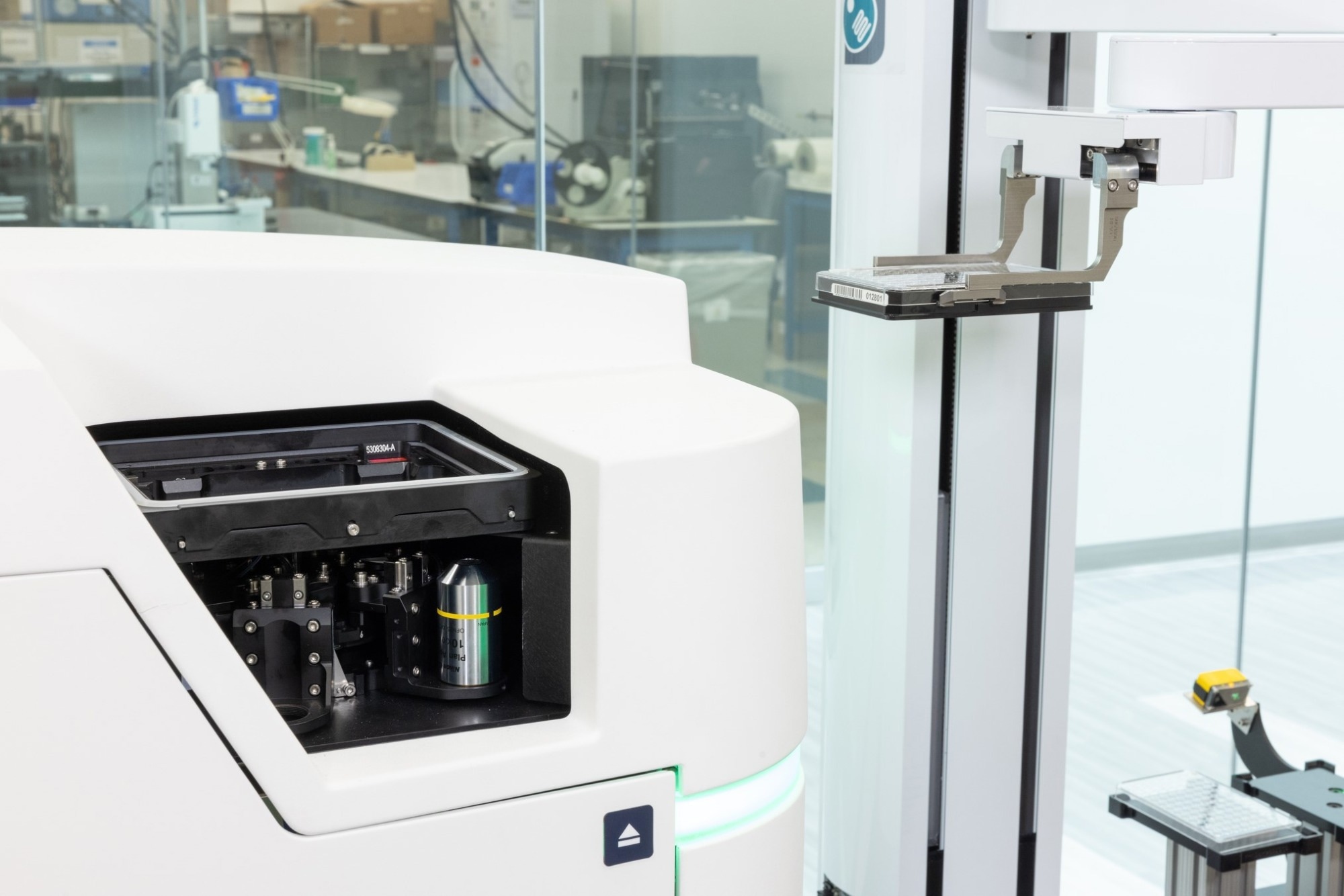
Robot-friendly loading design option for increased walk-away time with the ImageXpressHCS.ai High-Content Screening System. Image courtesy of Molecular Devices.
Where does AI improve image analysis most: Segmentation, classification, or quality control?
Segmentation is unquestionably the area where AI has the greatest impact.
Segmentation serves as the foundation for every subsequent analytical step. If segmentation is inaccurate, classification and quality control will also be compromised. Deep learning excels in situations where traditional threshold-based approaches struggle, particularly when dealing with complex or heterogeneous biological samples.
Once high-quality segmentation has been achieved, classification and quality control become more effective as they are built upon a reliable representation of the biological structures being analyzed.
In many ways, segmentation is the rate-limiting step in image analysis. Improving segmentation quality and speed creates a cascading benefit throughout the entire workflow, leading to better classification accuracy, stronger quality control, and faster analysis. As a result, AI-driven segmentation often delivers the greatest overall impact on high-content screening performance.
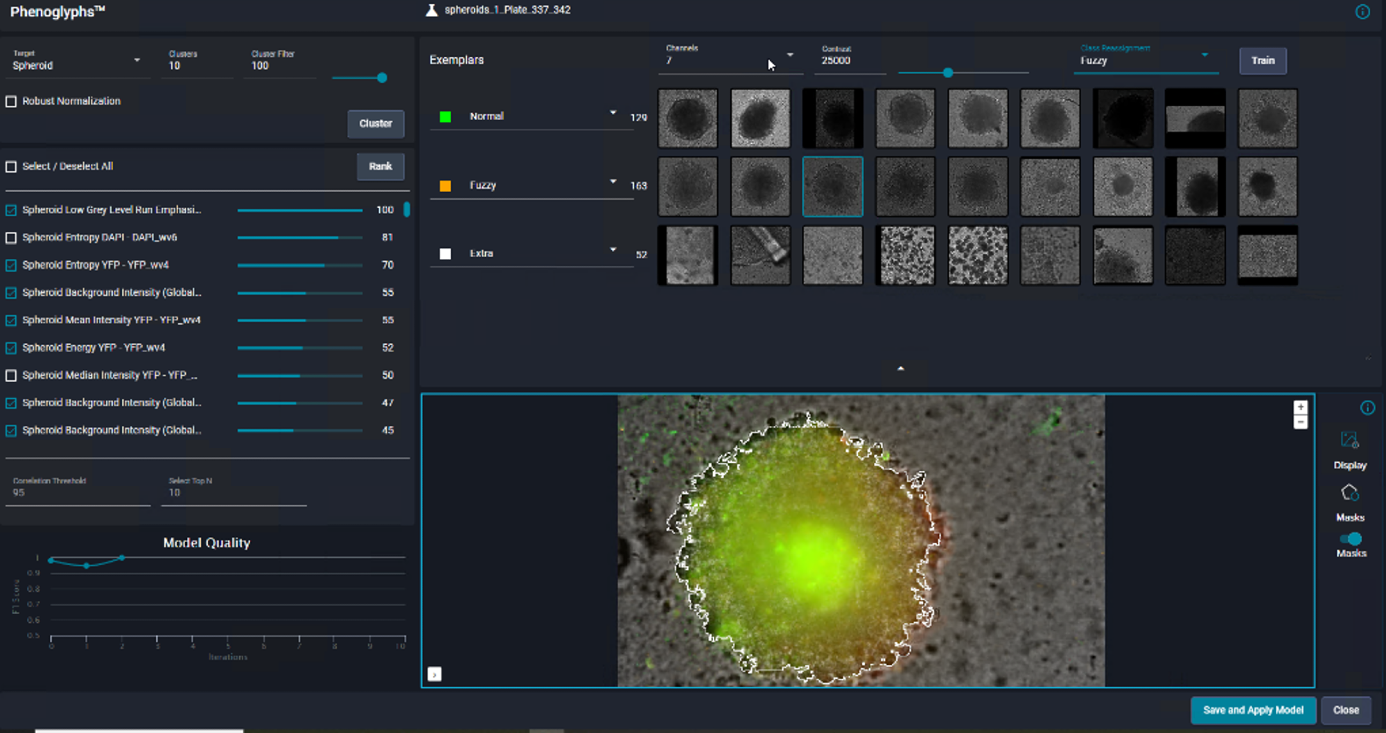
Classification of spheroids formed from HCT116 cells. Spheroids were segmented based on brightfield images using SINAP. Sample was additionally counter-stained with Hoechst 33342, Calcein AM and MitoTracker Red to visualize nuclei, live cells, and mitochondria, respectively. Image courtesy of Molecular Devices.
How do guided workflows help new users get reliable results faster?
One of the biggest bottlenecks in high-content screening is often protocol setup rather than image acquisition itself.
Guided workflows help eliminate this challenge by simplifying instrument configuration and protocol development. Instead of spending significant time building workflows from scratch, researchers can follow intuitive, step-by-step processes that accelerate setup and reduce errors.
This approach has two major benefits. First, it shortens the time between sample preparation and actionable results. Second, it improves reproducibility by allowing different users to follow the same standardized workflow and achieve comparable outcomes.
For core facilities and multi-user environments, guided workflows are especially valuable. They allow less experienced users to become productive quickly while ensuring that protocols remain consistent across users, projects, and locations.
How can machine learning reveal subtle phenotypic changes?
Machine learning excels at identifying patterns that are difficult or impossible for humans to detect consistently.
After segmentation has defined the relevant cellular structures, machine learning can evaluate numerous morphological features simultaneously, including cell shape, texture, spatial organization, organelle morphology, and many other parameters.
A researcher might visually assess only a handful of characteristics, but machine learning algorithms can analyze hundreds of features in parallel. This allows them to detect subtle phenotypic changes that would otherwise go unnoticed.
For example, in toxicology studies, compounds that appear harmless in traditional viability assays may still produce subtle changes in endoplasmic reticulum morphology, cytoskeletal organization, or nuclear architecture. Machine learning can identify these patterns objectively and classify them without human bias.
This capability is particularly powerful in approaches such as cell painting, where multiple cellular features are measured simultaneously to generate comprehensive phenotypic profiles. By detecting subtle changes earlier, machine learning helps researchers make better decisions and identify potential issues before they become more significant problems.
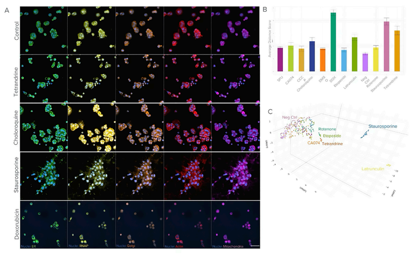
Cell painting assay on the ImageXpress HCS.ai System. A) Representative images of control and treated MCF7 cells. Scale=50µm. Images were analyzed using IN Carta® Image Analysis Software. The SINAP module was used for nuclei segmentation. 246 measurements per cell from the assay was uploaded to the StratoMineR™ software for further analysis. B) Graph showing the average physical distance score for compounds used. C) UMAP representation of the phenotypic profiles. Image courtesy of Molecular Devices.
How can core labs scale imaging capacity without adding staff?
For core facilities, time is often the most valuable resource.
Core laboratories support a diverse user base ranging from complete beginners to highly experienced researchers. A disproportionate amount of staff time is frequently spent on training, troubleshooting, and helping users navigate complex workflows.
AI-enabled imaging platforms and automated workflows help address this challenge by reducing the learning curve for new users. Researchers can become proficient much more quickly, allowing them to operate independently while still generating high-quality data.
As a result, core facility staff can focus on higher-value scientific activities such as experimental design, sample preparation strategies, and advanced data interpretation rather than routine operational support.
This enables facilities to support more users, run more experiments, and increase overall imaging capacity without expanding staffing levels. Improving usability, automation, and AI can effectively multiply the impact of existing personnel resources.
About Boyd Butler
Boyd Butler is a scientist and imaging specialist with extensive experience in microscopy, biophysics, and high-content screening. Throughout his career, he has served as a faculty member, principal investigator, and core facility director, supporting researchers across a wide range of biological and biomedical disciplines.
His academic training includes advanced study in biophysics and optical physics, providing a strong foundation in imaging technologies and quantitative image analysis. Over the years, he has worked extensively with microscopy platforms, image analysis workflows, and emerging AI-driven technologies designed to accelerate biological discovery.
At Molecular Devices, Butler focuses on helping scientists leverage advanced imaging solutions, including high-content screening systems, automated workflows, and AI-powered analytical tools. His expertise spans both the technical and practical aspects of imaging, from experimental design and sample preparation through to segmentation, classification, and phenotypic analysis.
Drawing on decades of experience in academia and core facility management, he is particularly passionate about improving accessibility to advanced imaging technologies, increasing reproducibility, and helping researchers generate meaningful biological insights more efficiently. His work continues to support the adoption of human-relevant model systems, machine learning-based image analysis, and next-generation workflows for drug discovery and translational research.
About Molecular Devices UK Ltd
Molecular Devices is one of the world’s leading providers of high-performance bioanalytical measurement systems, software and consumables for life science research, pharmaceutical and biotherapeutic development. Included within a broad product portfolio are platforms for high-throughput screening, genomic and cellular analysis, colony selection and microplate detection. These leading-edge products enable scientists to improve productivity and effectiveness, ultimately accelerating research and the discovery of new therapeutics. Molecular Devices is committed to the continual development of innovative solutions for life science applications. The company is headquartered in Silicon Valley, California, with offices around the globe. For more information, please visit www.moleculardevices.com.
 Making Complex Cell Culture Easy and Efficient with AI