Introduction
Home to billions of microorganisms of many different types, the human body has about an equal number of both human and non-human cells. These pathogens mainly include harmless bacteria, which can sometimes be helpful.
For instance, the biggest variety of species and the largest number of bacteria are present in the human gut than any other part of the body. Also known as enteric bacteria, the gut bacteria have a mutualistic association with the human body; these organisms grow by deriving nutrients from the human intestines, but they also aid in the digestion process and play a key role in the production of vitamins B and K.
However, these organisms may become harmful in certain cases, such as a weak immune system or during lack of hygiene, and induce mild to severe infections in other areas of the body. Bacteria can also form biofilms by aggregating in groups, and these biofilms adhere to one another and subsequently stick to surfaces, becoming more resistant toward antibiotics.
In fact, humans come into contact with biofilms on an everyday basis, with dental plaque being the most common biofilm. Dental plaque is predominantly formed by the bacteria Streptococcus mutans, but good oral hygiene can easily prevent this problem.
There are also other cases of biofilms that can be more harmful than the dental plaque. For instance, severe infections can be caused by biofilms that stick to medical appliances such as catheters or bones, and incidents such as these have raised concerns among many researchers.
Scanning electron microscopy
Researchers at the University Medical Center in Utrecht, examined an acute bone infection called osteomyelitis induced by Staphylococcus aureus1. This infection occurs when bacterial biofilm adheres together.
The study was carried out to develop a promising vaccine for this infection, and to achieve this, the team characterized proteins that are secreted by bacterial biofilms in vitro as well as human antibodies in vivo, and matched them to develop a potential vaccine.
The researchers then used a Scanning Electron Microscope (SEM) and successfully viewed the formation of biofilm on the human bone, together with cryo-SEM sections of the isolation of Staphylococcus aureus. This technique makes it easier to gain an insight into the various interactions that occur between the bones and the biofilm, and also between the bacteria themselves.
While a means to prevent biofilm formation is being explored by some researchers, another group at the Japan-based National Institutes of Natural Sciences is using Pseudomonas fluorescens2 to detect the genes involved in biofilm formation. The researchers initially detected a messenger-secreting gene known as c-di-GMP, which plays a role in the formation of biofilm.
They used a method based on transposons, which are sequences of DNA capable of changing their positions inside the genome. This allows them to alter genes and thus lead to silencing or over-expression of genes.
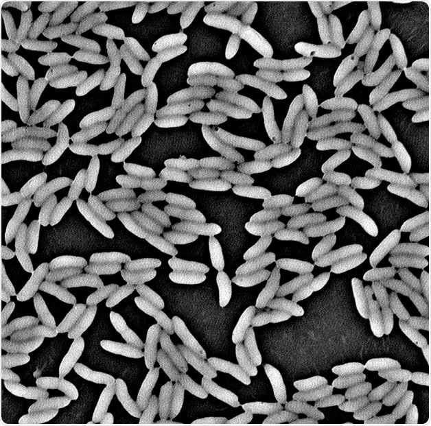
Figure 1. An example of the SEM image for the biofilm cells of P. fluorescens formed on a surface of polyvinyl chloride (magnification: 12,000x).
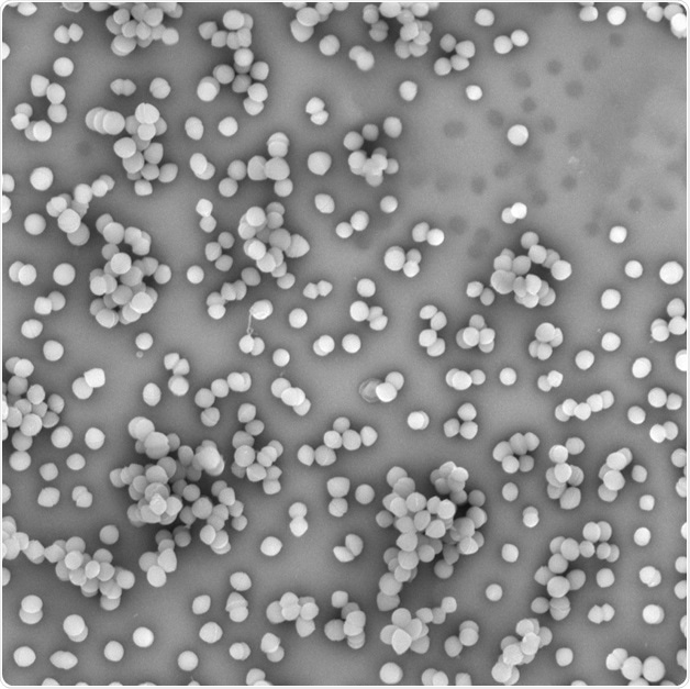
Figure 2. SEM image of bacteria Enterococcus faecium
Once the important gene was identified, it was muted and the biofilm forming capacities of the modified bacterial strain was compared against the normal, Wild Type strain. It was observed that the mutant-formed biofilm was reduced to less than 50% of the biofilm formed by the Wild Type strain, but there was no change in the cell number. This indicated that the reduction in biofilm size was in fact a reduction in the mutant bacterial cell size, and not due to its ability to multiply and produce a biofilm.
In order to further validate this theory, SEM imaging was performed on both strains. Then, using ParticleMetric software, the various sizes of bacteria were measured and a clear numerical comparison was made. Compared to the Wild Type strain, the mutant strain was found to be 25 to 30% smaller.
Conclusion
The size and shape of bacteria can often differ based on the environment, the presence of antibiotics, and the abundance of nutrients available. Here, the use of SEM in biofilm and microbiological research provides useful data about bacterial morphology. The technique also provides a clear picture of the microbiological realm in all of its states, thus driving the scope of research to the next level.
References
- Combining in vitro Protein Detection and in vivo Antibody Detection Identifies Potential Vaccine Targets Against Staphylococcus aureus During Osteomyelitis. den Reijer et al. Med Microbiol Immunol 2017 (2016)
- Disruption of de novo Purine Biosynthesis in Pseudomonas fluorescens Pf0-1 Leads to Reduced Biofilm Formation and a Reduction in Cell Size of Surface-Attached but not Planktonic Cells. Yoshioka et al. PeerJ (2016)
About Phenom-World

Phenom-World believe breakthroughs happen when complex nanotechnology is made intuitive, easier to use and brought within reach. As the leading global supplier of desktop scanning electron microscopes, their aim is to make imaging and analysis at the nanoscale available to every scientist in every lab.
That’s why Phenom-World invest their time and effort into developing high-quality electron microscope solutions that are functionally rich, yet simple to use. Because their mission is to create technology that has a real impact on how people work.
This takes innovative thinking, collaborative working and an entrepreneurial attitude. At their home in the high-tech region of Eindhoven in The Netherlands, Phenom-World have a group of passionate people who are given the freedom they need to innovate. Plus a highly skilled team of specialists in more than 40 countries who can provide support on a global scale.
Cooperation and co-creation are key to Phenom-World's success. They work closely with their partners to ensure they have the latest technologies, state-of-the-art equipment and applications for imaging and analysis. So they can get fast and accurate results and achieve more in nanotechnology.
Phenom-World is globally the yearly number 1 manufacturer of desktop scanning electron microscopes and imaging and analysis packages.
Sponsored Content Policy: News-Medical.net publishes articles and related content that may be derived from sources where we have existing commercial relationships, provided such content adds value to the core editorial ethos of News-Medical.Net which is to educate and inform site visitors interested in medical research, science, medical devices and treatments.