Sponsored Content by MerckReviewed by Emily MageeJan 10 2024
Tissue engineering and regenerative medicine hold great promise for enhancing and prolonging life, with multiple cell-based therapies now reaching the clinic.
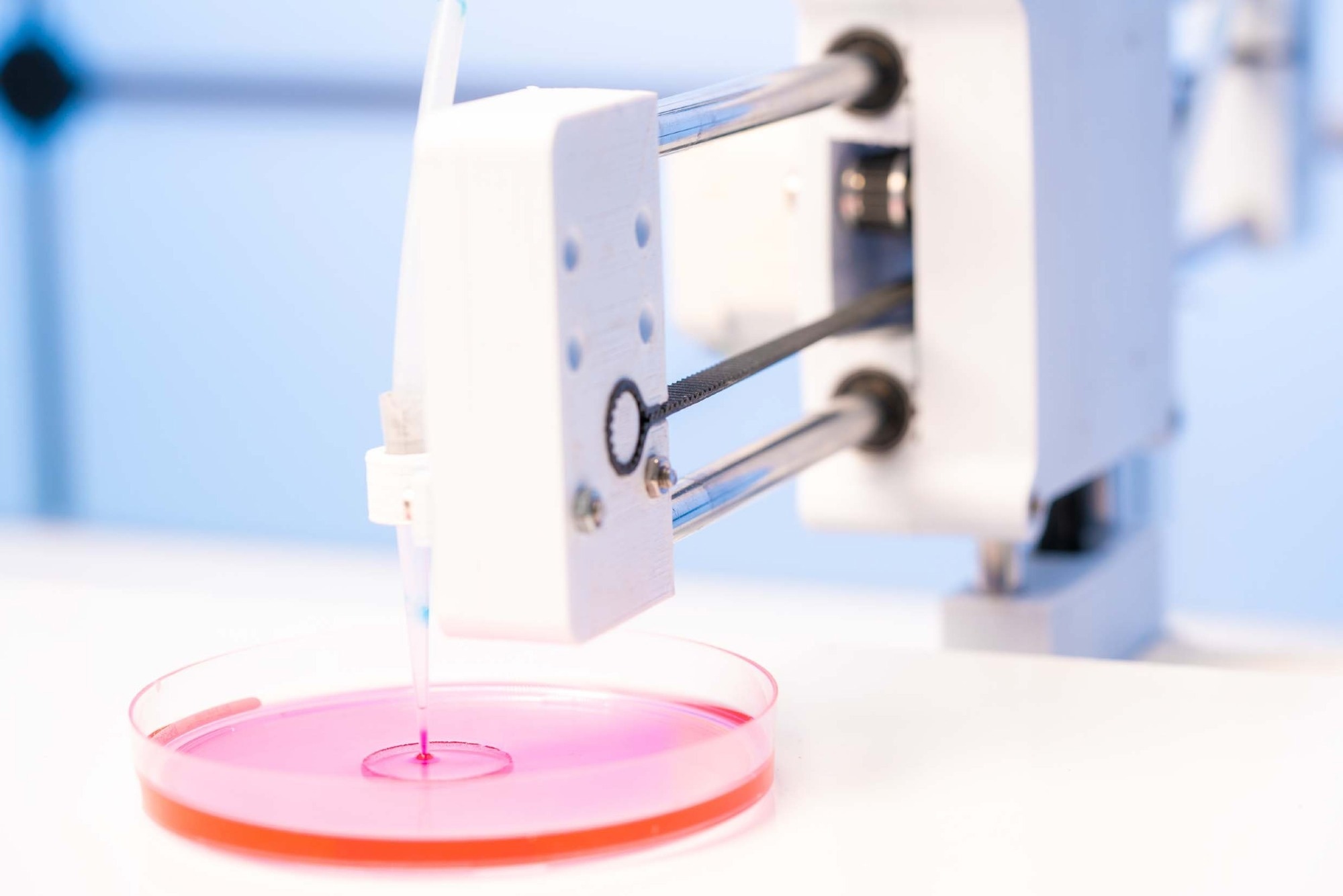
Image Credit: luchschenF/Shutterstock.com
From the replacement of entire organs to the delivery of therapeutic cells to an injured site, the efficacy of any tissue engineering strategy relies on the biomaterial scaffold utilized to deliver and/or develop cells in vitro and in vivo.1
Successful tissue engineering scaffolds have the following properties in common:
- They must be fabricated in a reproducible manner – this is part of the attractiveness of 3D printing strategies;2
- They must be easily delivered to the desired site of action, preferably through injection;1,3
- Ideally, they should match the mechanical and physical properties of the native tissue’s extracellular matrix (ECM);4
- They must support the desired cell functionality, such as proliferation, adhesion, and/or differentiation;5
- They should induce minimal or no inflammatory response;
- They must degrade at an optimal rate into nontoxic byproducts that are generally considered safe for clearance by the body.
Hydrogels are water-soluble polymer networks that are ideal for tissue engineering scaffolds due to their typically low inflammatory responses, high water content, highly variable chemistries, and tunable biomimetic mechanics.
A wide range of naturally occurring biopolymers such as proteins, polysaccharides, or nucleic acids (many of which may be found in the native ECM of cells) have been studied for utilization in tissue engineering applications.6
Typically, biopolymers display high cytocompatibility and can frequently degrade into safe extracellular matrix components such as amino acids and sugars.
However, the inherent bioactivity and the difficulty of reproducibly functionalizing such materials present challenges in controlling key properties such as degradation rate and mechanics.
Synthetic polymers can overcome these challenges because they can be engineered to create hydrogels with well-defined structures, chemistries, mechanics, and residual functionalities.
A synthetic approach enables the integration of multiple functional moieties, including some that have “smart” properties like in situ gelation, pH-responsiveness, and thermosensitivity, using typically facile copolymerization strategies.4,7
In some instances, synthetic polymers can also be modified to degrade into small molecules. For example, the degradable polymer poly(lactide-co-glycolide) (PLGA) can degrade into natural metabolites like glycolic and lactic acid.
However, there are fewer examples of water-soluble hydrogel precursor polymers with this functionality.4 Alternatively, synthetic hydrogels can be designed to allow clearance by the renal system by controlling the polydispersity and polymer size and linking the polymers with degradable crosslinks.3
Poly (ethylene glycol) (PEG) is the most extensively studied synthetic polymer for tissue engineering. PEG provides benefits, including a hydrated structure, which typically leads to high cytocompatibility and protein repellency to reduce inflammatory responses.8
The hydrophilicity of PEG means that it is usually non-adhesive to cells. However, the incorporation of cellular adhesive motifs (e.g., arginylglycylaspartic acid (RGD)) within the hydrogel helps to overcome this issue.7
Based on these properties, PEG-based hydrogels have been successfully applied to a range of tissue engineering and cell delivery applications.9 However, conventional PEG-based hydrogels are limited in terms of their lack of injectability, lack of tunable degradation, and relatively poor mechanics.
This article will outline how changing the crosslinking chemistry and/or the chain structure of the PEG starting materials can enhance the performance of PEG and PEG-derivative hydrogels for tissue engineering or cell delivery applications.
Crosslinking chemistry
The most widely utilized strategy for the preparation of PEG-based hydrogels is the free radical polymerization of poly(ethylene glycol) diacrylates, with the degree of crosslinking modified by changing the length of the PEG chain between the acrylate groups.10
However, the lack of injectability, non-degradability, low swelling ratio, and uncontrolled mesh size of the resultant hydrogels can produce limitations in biomedical applications. As a result, alternative crosslinking strategies have been developed to offer both degradability and injectability in PEG-based networks.
Injectability
The most widely employed strategy to produce injectable PEG-based hydrogels is the co-extrusion of two PEG derivatives functionalized with complementary groups that can either chemically or physically crosslink.
Covalent in situ-gelation, the more common of the two in tissue engineering, is enabled by converting the hydroxyl end group of PEG to a variety of rapidly reactive functional groups to enable one or more various in situ crosslinking chemistries.
Controlling the length, number, and reversibility of crosslinks enables the physical properties of a hydrogel to be tuned.3
While the application of these chemistries to different cell scaffolding applications has been successful, each brings its own advantages and disadvantages. Michael addition chemistry is relatively quick but results in a non-degradable linkage and may interact with proteins in the body.
Disulfide formation produces redox-responsive hydrogels but is usually slower, and the resultant gels are typically weaker.
Hydrazone chemistry is rapid and produces a hydrolyzable hydrazone bond but typically utilizes aldehyde groups that can create Schiff bases with amines in proteins.
Oxime chemistry results in a bond that is more slowly degradable, and this is suitable for longer-lasting scaffolds. However, this process demands acid catalysis for rapid gelation.
Diels-Alder chemistry is highly bio-orthogonal, but gels relatively slowly and leads to functionally irreversible crosslinks (at least without high temperatures).
Strain-promoted alkyne/azide cycloaddition is rapid and highly specific but causes the hydrogel to be hydrophobic.
As a result, the crosslinking chemistry must be carefully selected to balance the potential for side-reactions, the gelation rate, and the rate of degradation that is most appropriate for each application.
Non-covalent interactions, including hydrogen bonding, stereocomplexation, electrostatics, or hydrophobic interactions, can also be employed to facilitate in situ gelation or to produce highly shear-thinning hydrogels that still allow injection.11
However, these strategies frequently result in hydrogels that have poor stability in the highly diluted environment of the body and/or are subject to interferences in vivo that interrupt crosslinking.
Yet, inclusion complexes prepared using cyclic supramolecular structures like alpha-cyclodextrin (CD) as a host for polymers such as poly (ethylene glycol) do offer an interesting alternative to covalent crosslinking.12
Multiple PEG chains can thread through the center of the CD and interact via dipole interactions/hydrogen bonding to form a crosslinking point. The mobility of the inclusion complex crosslinks (unlike covalent crosslinks) is relevant to tissue engineering as it can result in self-healing hydrogel structures.
Using a combination of covalent and noncovalent crosslinking strategies offers interesting options that can overcome the disadvantages of both physical and covalent approaches.
For instance, Qiao et al. successfully functionalized PEG using alkyne end-groups and CD rings with reactive azide groups to facilitate both covalent (alkyne/azide click chemistry) and non-covalent (CD-PEG interactions) crosslinking.
Both HEK293T and HeLa cells could be maintained inside the scaffold with over 90% viability in 10 days, and the gel can experience complete degradation within a month.13
Degradability
To allow optimal tissue regeneration, the degradation rate of a scaffold must match the rate at which the cells propagate through the matrix and form their own extracellular matrix, recreating native biology.
Although common degradation mechanisms such as hydrolysis can be engineered to enable such control, the use of specific stimuli such as light or disease state to dynamically degrade the gel under specific environmental conditions in which degradation is desirable is highly attractive.5
The most popular approach is enzymatic degradation, which involves specific amino acid sequences that are substrates to various enzymes (including those specifically up-regulated in disease) being employed as crosslinkers.
The most reported substrate is matrix metalloproteases (MMPs), cell-secreted enzymes that are overexpressed in several inflammatory conditions and cancers.7
For instance, Bryant et al. reported a PEG-based hydrogel that was loaded with pre-hydroxyapatite nanoparticles and osteoblast cells and crosslinked with MMP-sensitive peptide sequences; the degradation rate of the hydrogel was accelerated upon the upregulation of MMPs during osteogenesis to promote bone regeneration.9
Additionally, visible or UV light has been employed to induce targeted degradation in hydrogels. Crosslinks may be manipulated using light-induced reversible ring-opening/ring-closing isomerization (e.g., diarylethenes, spiropyrans, fulgimides) or cis-trans isomerization (e.g., azobenzenes or stilbens).5
Despite this approach being largely limited to in vitro use because of the low penetration of light through the body, it can also be employed in in vivo dental, topical, and optical implants to initiate photoinduced degradation to degrade or reduce the stiffness of the gel.
The latter is useful for promoting cell proliferation and inducing specific cell differentiation.
For instance, Anseth’s group created a cellularized hydrogel by crosslinking thiolated 4-armed star PEG and acrylate functionalized poly(ethylene glycol) di-photodegradable acrylate (PEGdiPDA). This was carried out using Michael addition chemistry and the subsequent degradation of the hydrogel using cleavage of the o-nitrobenzyl ether photodegradable linker via UV irradiation.14
The on-demand switching of the hydrogel mechanics and porosity allowed the precise control of cellular responses, including single stem cell migration15 and cell differentiation (such as the transition from fibroblast to myofibroblast).16
Chain structure
The chemical structure of PEG and PEG-based derivatives serves a vital function in the regulation of the biological responses, hydrophilicity, protein repellency, degradation kinetics, mechanical properties, and swelling of PEG-based hydrogels.17
A popular modification is the attachment of biodegradable hydrophobic polymer blocks based on polymers like poly (lactic acid) (PLA), poly(propylene oxide) (PPO), or poly (lactide-co-glycolide) (PLGA) to PEG to create diblock or triblock copolymers.
These copolymers can self-associate into hydrogels through hydrophobic interactions, but degradation occurs over time as the hydrophobic polymer also degrades.18
Triblock copolymers, particularly PEO-PPO-PEO (i.e., the Pluronics family of polymers), have been frequently studied due to the sol-gel transition behavior that they exhibit upon heating from room temperature to physiological temperature, and this facilitates injection.
For example, Kolesky et al. reported the utilization of PEO-PPO-PEO as a thermogelling cell-laden ink to produce vascularized tissue constructs that contain fibroblast cells via bioprinting.19 These networks are susceptible to dilution over time and have relatively poor mechanics.
These factors limit such materials to being used in lower modulus tissue engineering, although combining these polymers with covalent bond-forming units (such as diacrylated Pluronic F-127)20 can partially overcome these issues.
Additionally, changing the physical arrangement of the PEG chains from linear chains to more complex geometries has a substantial effect on the gel properties.
Branched or star arrangements of PEG chains allow the formation of higher modulus and chemically tunable PEG hydrogels due to the increasing number of crosslinkable groups per single chain in comparison to linear PEG, which has only one –OH group at each end of the chain.21
Spatially organizing PEG chains in the precursor materials also enables the creation of more ordered and well-defined chemical structures that are advantageous to the promotion of desirable cell responses.22
The preparation of star PEG polymers typically involves the connection of 4, 6 or 8 arms of linear PEG (of a tunable length) to a single internal point; the end group of each arm may be functionalized using crosslinkers or biomolecules as required to yield gels with more tunable mechanics and biological activities.23
Tetrahedron-oriented 4-arm star PEGs have also been proven to create highly homogeneous network structures, allowing the production of hydrogels with moduli in the MPa range.24 Additionally, star-PEGs may be employed in combination with injectable or degradable chemistries to produce tissue scaffolds.
For example, amphiphilic 8-armed PEG-b-PLA-cholesterol copolymers thermally gel at 34 °C because of interactions between the cholesterol groups to create a microstructured network that can support L929 mouse fibroblast cell viability and proliferation in the hydrogel.25
Utilizing the extra surface functionality of star PEGs enables the production of highly functional hydrogels without compromising the crosslinking density.
For example, RGD-modified star PEG coatings can considerably improve cell spreading in comparison to non-RGD functionalized star PEG or RGD-functionalized linear PEG gels due to the higher density of RGD that may be grafted with a star-PEG morphology.26
To prevent the multiple-step synthesis of star PEGs, hyperbranched PEG copolymers (globular chains with molecular weights between 1400 g/mol and 1,700,000 g/mol) can be synthesized using the random anionic ring-opening copolymerization of ethylene oxide in the presence of glycidol.27
This straightforward free radical reaction technique for the production of a PEG derivative with a high number of functionalizable chain ends is practically and synthetically attractive despite it producing a more polydisperse starting unit than star PEGs (resulting in weaker gels) and possibly leading to low yields.
It is believed that such materials have not yet been studied in-depth for tissue engineering applications.
Branched PEG copolymers provide an alternative and highly attractive option for creating PEG-based hydrogels. The most common branched PEG materials are based on poly(oligoethylene glycol methacrylate) (POEGMA), which are comprised of a methacrylate backbone and one PEG side chain of tunable length per monomer repeat unit.17
Such a structure offers facile polymerization via free or controlled radical processes, enabling the formation of both linear and hyperbranched structures as well as easy functionalizability by copolymerization.28 Additionally, adjusting the length of the PEG side chain significantly alters the hydrogel’s properties.
For example, Lutz and coworkers demonstrated that copolymerization of long-side chain oligo (ethylene glycol) methyl ether methacrylate (OEGMA) and short-side chain di(ethylene glycol) methyl ether methacrylate (M(EO)2MA) monomers leads to polymers and hydrogels with a precisely tunable lower critical solution temperature (LCST) between room temperature to > 80 °C according to the ratio of OEGMA and M(EO)2MA used.
LCST behavior changes the hydrogel from being highly hydrophilic to being relatively hydrophobic, driving considerable changes in cell adhesion and tunable cell delamination.30
Merck’s laboratories have actively developed printable or injectable analogs of POEGMA hydrogels through the functionalization of POEGMA precursor polymers using aldehyde and hydrazide reactive functional groups. This allows in situ gelation to occur after mixing-based coextrusion takes place using hydrazone crosslinking.31
Adjusting the molecular weight, concentration, or density of the reactive functional groups on the precursor polymers allows the gelation time to be adjusted from minutes to hours for specific applications.
This enables facile injection in vivo, in addition to processing via methods such as electrospinning, to produce well-defined nanofibrous hydrogel networks with the potential to simulate the nanofibrous extracellular matrix32 or inkjet printing to produce immobilized hydrogel coatings on porous supports.33
The PEG-based chemistry maintains low inflammatory responses upon injection in vivo, while the hydrazone gelation chemistry enables hydrolytic degradation to occur over time. It has been demonstrated that branched PEGs can prevent some of the emerging immune responses observed with PEG-based materials.34
Additionally, the facile copolymerizability of POEGMA allows the development of a range of functional POEGMA-based copolymers that are injectable while introducing the potential for temperature-responsive swelling, hydrophobic domain formation, cell-adhesive properties, or cell delamination.17
From a cell delivery perspective, introducing charged groups via the copolymerization of POEGMA with N,N-dimethylaminoethyl methacrylate (DMAEMA, cationic charge) or acrylic acid (AA, anionic charge) resulted in significantly improved cell adhesion.
Amphoteric hydrogels that were prepared by mixing anionic and cationic-functionalized precursor polymers offered enhanced cell viability for the long-term encapsulation and delivery of retinal pigment epithelial cells to the back of the eye.35
Conclusion
The bio-inert nature of PEG has enabled PEG hydrogels to be utilized effectively in a range of cell delivery and tissue engineering applications.
The tunable chemical properties of PEG derivatives, such as star/branched and injectable PEGs, hold great promise when it comes to controlling the degradability, chemistry, mechanics, and injectability of PEG-based hydrogels to enhance their performance in cell-based applications.
Merck believes that the considerably enhanced chemical flexibility of PEG derivative hydrogels can overcome the challenges of conventional PEG hydrogels, especially regarding the improvement of the mechanics and cell-hydrogel interactions to encourage desired tissue growth or cell differentiation/maintenance responses.
Acknowledgments
Produced from materials originally authored by Ali Affar, Fei Xu, and Todd Hoare
From McMaster University, Department of Chemical Engineering, Ontario, Canada.
References
- Liu M, Zeng X, Ma C, Yi H, Ali Z, Mou X, Li S, Deng Y, He N. 2017. Injectable hydrogels for cartilage and bone tissue engineering. Bone Res. 5(1): https://doi.org/10.1038/boneres.2017.14
- Kang H, Lee SJ, Ko IK, Kengla C, Yoo JJ, Atala A. 2016. A 3D bioprinting system to produce human-scale tissue constructs with structural integrity. Nat Biotechnol. 34(3):312-319. https://doi.org/10.1038/nbt.3413
- Patenaude M, Smeets NMB, Hoare T. 2014. Designing Injectable, Covalently Cross-Linked Hydrogels for Biomedical Applications. Macromol. Rapid Commun.. 35(6):598-617. https://doi.org/10.1002/marc.201300818
- Khan F, Tanaka M. Designing Smart Biomaterials for Tissue Engineering. IJMS. 19(1):17. https://doi.org/10.3390/ijms19010017
- Brown TE, Anseth KS. Spatiotemporal hydrogel biomaterials for regenerative medicine. Chem. Soc. Rev.. 46(21):6532-6552. https://doi.org/10.1039/c7cs00445a
- Bedian L, Villalba-Rodríguez AM, Hernández-Vargas G, Parra-Saldivar R, Iqbal HM. 2017. Bio-based materials with novel characteristics for tissue engineering applications ? A review. International Journal of Biological Macromolecules. 98837-846. https://doi.org/10.1016/j.ijbiomac.2017.02.048
- Lu Y, Aimetti AA, Langer R, Gu Z. 2017. Bioresponsive materials. Nat Rev Mater. 2(1): https://doi.org/10.1038/natrevmats.2016.75
- Khoushabi A, Schmocker A, Pioletti D, Moser C, Schizas C, Månson J, Bourban P. 2015. Photo-polymerization, swelling and mechanical properties of cellulose fibre reinforced poly(ethylene glycol) hydrogels. Composites Science and Technology. 11993-99. https://doi.org/10.1016/j.compscitech.2015.10.002
- Carles-Carner M, Saleh LS, Bryant SJ. The effects of hydroxyapatite nanoparticles embedded in a MMP-sensitive photoclickable PEG hydrogel on encapsulated MC3T3-E1 pre-osteoblasts. Biomed. Mater.. 13(4):045009. https://doi.org/10.1088/1748-605x/aabb31
- Tan S, Blencowe A, Ladewig K, Qiao GG. 2013. A novel one-pot approach towards dynamically cross-linked hydrogels. Soft Matter. 9(21):5239. https://doi.org/10.1039/c3sm50638j
- Zhu J. 2010. Bioactive modification of poly(ethylene glycol) hydrogels for tissue engineering. Biomaterials. 31(17):4639-4656. https://doi.org/10.1016/j.biomaterials.2010.02.044
- Wylie RG, Ahsan S, Aizawa Y, Maxwell KL, Morshead CM, Shoichet MS. 2011. Spatially controlled simultaneous patterning of multiple growth factors in three-dimensional hydrogels. Nature Mater. 10(10):799-806. https://doi.org/10.1038/nmat3101
- Sawhney AS, Pathak CP, Hubbell JA. 1993. Bioerodible hydrogels based on photopolymerized poly(ethylene glycol)-co-poly(.alpha.-hydroxy acid) diacrylate macromers. Macromolecules. 26(4):581-587. https://doi.org/10.1021/ma00056a005
- Revzin A, Russell RJ, Yadavalli VK, Koh W, Deister C, Hile DD, Mellott MB, Pishko MV. 2001. Fabrication of Poly(ethylene glycol) Hydrogel Microstructures Using Photolithography. Langmuir. 17(18):5440-5447. https://doi.org/10.1021/la010075w
- Wang Y, Adokoh CK, Narain R. 2018. Recent development and biomedical applications of self-healing hydrogels. Expert Opinion on Drug Delivery. 15(1):77-91. https://doi.org/10.1080/17425247.2017.1360865
- Poudel AJ, He F, Huang L, Xiao L, Yang G. 2018. Supramolecular hydrogels based on poly (ethylene glycol)-poly (lactic acid) block copolymer micelles and ?-cyclodextrin for potential injectable drug delivery system. Carbohydrate Polymers. 19469-79. https://doi.org/10.1016/j.carbpol.2018.04.035
- Tan S, Blencowe A, Ladewig K, Qiao GG. 2013. A novel one-pot approach towards dynamically cross-linked hydrogels. Soft Matter. 9(21):5239. https://doi.org/10.1039/c3sm50638j
- Kloxin AM, Kasko AM, Salinas CN, Anseth KS. 2009. Photodegradable Hydrogels for Dynamic Tuning of Physical and Chemical Properties. Science. 324(5923):59-63. https://doi.org/10.1126/science.1169494
- Tibbitt, M. W.; Kloxin, A. M.; Sawicki, L. A.; Anseth, K. S. . Macromolecules2013, 46, 2785..
- Guo Q, Wang X, Tibbitt MW, Anseth KS, Montell DJ, Elisseeff JH. 2012. Light activated cell migration in synthetic extracellular matrices. Biomaterials. 33(32):8040-8046. https://doi.org/10.1016/j.biomaterials.2012.07.013
- Kloxin AM, Benton JA, Anseth KS. 2010. In situ elasticity modulation with dynamic substrates to direct cell phenotype. Biomaterials. 31(1):1-8. https://doi.org/10.1016/j.biomaterials.2009.09.025
- Bakaic E, Smeets NMB, Hoare T. Injectable hydrogels based on poly(ethylene glycol) and derivatives as functional biomaterials. RSC Adv.. 5(45):35469-35486. https://doi.org/10.1039/c4ra13581d
- Jeong B, Bae YH, Kim SW. 1999. Thermoreversible Gelation of PEG?PLGA?PEG Triblock Copolymer Aqueous Solutions. Macromolecules. 32(21):7064-7069. https://doi.org/10.1021/ma9908999
- Zhu KJ, Xiangzhou L, Shilin Y. 1990. Preparation, characterization, and properties of polylactide (PLA)?poly(ethylene glycol) (PEG) copolymers: A potential drug carrier. J. Appl. Polym. Sci.. 39(1):1-9. https://doi.org/10.1002/app.1990.070390101
- Kolesky DB, Truby RL, Gladman AS, Busbee TA, Homan KA, Lewis JA. 2014. 3D Bioprinting of Vascularized, Heterogeneous Cell-Laden Tissue Constructs. Adv. Mater.. 26(19):3124-3130. https://doi.org/10.1002/adma.201305506
- Yoon JJ, Chung HJ, Park TG. 2007. Photo-crosslinkable and biodegradable Pluronic/heparin hydrogels for local and sustained delivery of angiogenic growth factor. J. Biomed. Mater. Res.. 83A(3):597-605. https://doi.org/10.1002/jbm.a.31271
- Keys KB, Andreopoulos FM, Peppas NA. 1998. Poly(ethylene glycol) Star Polymer Hydrogels. Macromolecules. 31(23):8149-8156. https://doi.org/10.1021/ma980999z
- Inoue K. 2000. Functional dendrimers, hyperbranched and star polymers. 25(4):453-571. https://doi.org/10.1016/s0079-6700(00)00011-3
- Freudenberg U, Hermann A, Welzel PB, Stirl K, Schwarz SC, Grimmer M, Zieris A, Panyanuwat W, Zschoche S, Meinhold D, et al. 2009. A star-PEG?heparin hydrogel platform to aid cell replacement therapies for neurodegenerative diseases. Biomaterials. 30(28):5049-5060. https://doi.org/10.1016/j.biomaterials.2009.06.002
- Sakai T, Matsunaga T, Yamamoto Y, Ito C, Yoshida R, Suzuki S, Sasaki N, Shibayama M, Chung U. 2008. Design and Fabrication of a High-Strength Hydrogel with Ideally Homogeneous Network Structure from Tetrahedron-like Macromonomers. Macromolecules. 41(14):5379-5384. https://doi.org/10.1021/ma800476x
- Nagahama K, Ouchi T, Ohya Y. 2008. Temperature-Induced Hydrogels Through Self-Assembly of Cholesterol-Substituted Star PEG-b-PLLA Copolymers: An Injectable Scaffold for Tissue Engineering. Adv. Funct. Mater.. 18(8):1220-1231. https://doi.org/10.1002/adfm.200700587
- Groll J, Fiedler J, Engelhard E, Ameringer T, Tugulu S, Klok H, Brenner RE, Moeller M. 2005. A novel star PEG-derived surface coating for specific cell adhesion. J. Biomed. Mater. Res.. 74A(4):607-617. https://doi.org/10.1002/jbm.a.30335
- Perevyazko I, Seiwert J, Schömer M, Frey H, Schubert US, Pavlov GM. 2015. Hyperbranched Poly(ethylene glycol) Copolymers: Absolute Values of the Molar Mass, Properties in Dilute Solution, and Hydrodynamic Homology. Macromolecules. 48(16):5887-5898. https://doi.org/10.1021/acs.macromol.5b01020
- Wilms D, Schömer M, Wurm F, Hermanns MI, Kirkpatrick CJ, Frey H. 2010. Hyperbranched PEG by Random Copolymerization of Ethylene Oxide and Glycidol. Macromol. Rapid Commun.. 31(20):1811-1815. https://doi.org/10.1002/marc.201000329
- Luzon M, Boyer C, Peinado C, Corrales T, Whittaker M, Tao L, Davis TP. 2010. Water-soluble, thermoresponsive, hyperbranched copolymers based on PEG-methacrylates: Synthesis, characterization, and LCST behavior. J. Polym. Sci. A Polym. Chem.. 48(13):2783-2792. https://doi.org/10.1002/pola.24027
- Lutz J, Andrieu J, Üzgün S, Rudolph C, Agarwal S. 2007. Biocompatible, Thermoresponsive, and Biodegradable: Simple Preparation of ?All-in-One? Biorelevant Polymers. Macromolecules. 40(24):8540-8543. https://doi.org/10.1021/ma7021474
- Lutz J, Akdemir Ö, Hoth A. 2006. Point by Point Comparison of Two Thermosensitive Polymers Exhibiting a Similar LCST: Is the Age of Poly(NIPAM) Over?. J. Am. Chem. Soc.. 128(40):13046-13047. https://doi.org/10.1021/ja065324n
- Lutz J. 2011. Thermo-Switchable Materials Prepared Using the OEGMA-Platform. Adv. Mater.. 23(19):2237-2243. https://doi.org/10.1002/adma.201100597
- Wischerhoff E, Uhlig K, Lankenau A, Börner H, Laschewsky A, Duschl C, Lutz J. 2008. Controlled Cell Adhesion on PEG-Based Switchable Surfaces. Angewandte Chemie International Edition. 47(30):5666-5668. https://doi.org/10.1002/anie.200801202
- Bakaic E, Smeets NMB, Dorrington H, Hoare T. ?Off-the-shelf? thermoresponsive hydrogel design: tuning hydrogel properties by mixing precursor polymers with different lower-critical solution temperatures. RSC Adv.. 5(42):33364-33376. https://doi.org/10.1039/c5ra00920k
- Smeets NMB, Bakaic E, Patenaude M, Hoare T. 2014. Injectable and tunable poly(ethylene glycol) analogue hydrogels based on poly(oligoethylene glycol methacrylate). Chem. Commun.. 50(25):3306. https://doi.org/10.1039/c3cc48514e
- Xu F, Sheardown H, Hoare T. Reactive electrospinning of degradable poly(oligoethylene glycol methacrylate)-based nanofibrous hydrogel networks. Chem. Commun.. 52(7):1451-1454. https://doi.org/10.1039/c5cc08053c
- Mateen R, Ali MM, Hoare T. 2018. A printable hydrogel microarray for drug screening avoids false positives associated with promiscuous aggregating inhibitors. Nat Commun. 9(1): https://doi.org/10.1038/s41467-018-02956-z
- Liu M, Johansen P, Zabel F, Leroux J, Gauthier MA. 2014. Semi-permeable coatings fabricated from comb-polymers efficiently protect proteins in vivo. Nat Commun. 5(1): https://doi.org/10.1038/ncomms6526
- (a) Bakaic, E.; Smeets, N. M. B.; Badv, M.; Dodd, M.; Barrigar, O.; Siebers, E.; Lawlor, M.; Sheardown, H.; Hoare, T. ACS Biomater. Sci. Eng.2017, DOI: 10.1021/acsbiomaterials.7b00397.
- (b) Bakaic, E.; Smeets, N. M. B.; Barrigar, O.; Alsop, R.; Rheinstädter, M. C.; Hoare, T. Macromolecules2017,50, 7687.
About Merck
Our pursuit is progress for people everywhere. That's why we take a closer look at things, ask questions, and think ahead.
We've been around for more than 350 years, yet our majority owners are still the descendants of Friedrich Jacob Merck, the man who founded our company in Darmstadt, Germany in 1668.
From advancing gene-editing technologies and discovering unique ways to treat the most challenging diseases to enabling the intelligence of devices – the company is everywhere.
We are Merck. The only exceptions are the United States and Canada. Here we operate as EMD Serono in the Biopharma business, as MilliporeSigma in the Life Science business, and as EMD Performance Materials in the materials business.
Our life science business
We provide infinite solutions to solve the toughest problems in life science in collaboration with the global scientific community. Our tools, services, and digital platforms empower scientists and engineers at every stage, helping deliver breakthrough therapies more quickly.
Focus areas
With our three business units, we are a leading worldwide supplier of tools, high-grade chemicals, and equipment for academic labs, biotech, and biopharmaceutical manufacturers, as well as the industrial sector.
- Research Solutions provides our academic customers with the chemicals and tools needed to make scientific discovery easier and faster.
- Process Solutions provides drug manufacturers with process development expertise and technologies, such as continuous bioprocessing.
- Applied Solutions offers both testing kits and services to ensure that our food is safe to eat and our water is clean to drink.
Sponsored Content Policy: News-Medical.net publishes articles and related content that may be derived from sources where we have existing commercial relationships, provided such content adds value to the core editorial ethos of News-Medical.Net which is to educate and inform site visitors interested in medical research, science, medical devices and treatments.