The automated CellHesion® 300 is the perfect instrument for quantifying cell-tissue, cell-cell, and cell-substrate communications with the sensitivity of a single molecule. It allows quick and effortless measurement of the morphology, structure, and nanomechanical characteristics of living biological systems, offering a key understanding of their role in different pathological disorders.
This advanced system develops new prospects for applications in biochemistry, biophysics, wound healing, implant research, developmental biology, infection biology, stem cell research, and immune response studies.
Features
Understanding biomechanics made easy
CellHesion 300 offers reproducible, quantitative data of exceptional quality. Its great level of automation enhances throughput and provides the performance, productivity, and statistical importance necessary for clinical and biomedical research environments. Significant factors, such as utmost adhesion force, discrete unbinding events, and tether features, are automatically determined in every dataset by advanced software solutions.
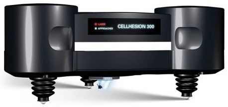
Image Credits: Bruker Nano GmbH - JPK BioAFM.
CellHesion 300 delivers:
- In near-physiological conditions, label-free and correlative multiparametric, nanomechanical measurements of living samples
- For increased productivity, maximized throughput by automating measurements
- Systematic workflow with visual support
- Simple and quick selection of areas of interest over huge sample regions, suitable for tissue biopsies
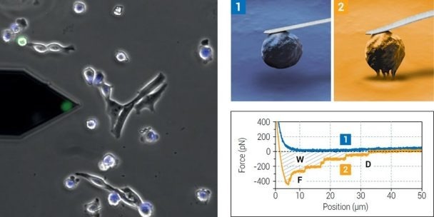
Left: Probe with a single 3T3 fibroblast (green, FDA staining) attached for cell adhesion measurements on a substrate or target cells. Right: In a Single Cell Force Spectroscopy (SCFS) experiment, a single living cell is biochemically bound to a probe (e.g., via functionalization). The cell is brought into contact with the binding target and a defined force applied to the cell (1). After a user-defined binding period, the cell is separated from target by retracting the probe (2). The resistance to separation is measured by quantification of the probe deflection. Image Credits: Bruker Nano GmbH - JPK BioAFM.
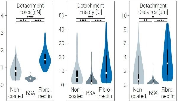
Violin plots showing the distribution of Detachment Energy (W), Force (F), and Distance (D), determined by Single Cell Force Spectroscopy detachment measurements between a single 3T3 mouse fibroblast and a polyacrylamide hydrogel (50kPa) with different coatings. Sample courtesy of Dr. Stephanie Wedepohl, Freie Universität Berlin, Germany. Image Credits: Bruker Nano GmbH - JPK BioAFM
Automatic mechanical screening of highly topographic samples with the CellHesion 300 is of great advantage for tissue characterization in the clinical context.”
Prof. Dr. Ansgar Petersen, BIH, Center for Regenerative Therapies, Charité Medical University
Performing automated multi-parametric nanomechanical mapping
State-of-the-art software, automated alignment, and intuitive user guidance of the detection system pave the way for self-directed operation and quick results. The novel CellHesion 300 user interface offers fast and effortless control of the system and its parameters.
For user-defined experiments, the software provides a practical scripting tool. Efficient batch data processing capacities offer the quantification and analysis of huge datasets at a single button touch.
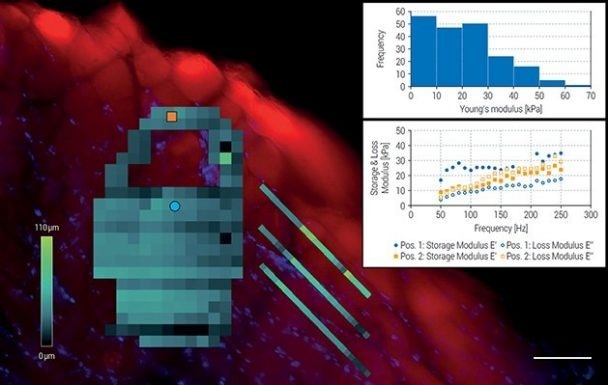
Force maps of user-defined shapes on a cross-section of sheep muscle tissue overlayed with an optical fluorescence tiling image (scalebar 100 µm). Muscle fibers, rich in actin filaments, were stained with phalloidin-TRITC (red) and cell nuclei with DAPI (blue). Internal dark areas depict unlabeled connective tissue. Force maps were acquired in SmartMapping mode and illustrate the combined height measurement of samples with large topographies. Upper inset: Distribution of Young’s modulus values. Lower inset: Plot of storage modulus and loss modulus at 2 different positions on the tissue sample (blue circle and orange square). Sheep muscle sample also used for cover image. Sample courtesy of Prof. Dr. Ansgar Petersen, BIH, Center for Regenerative Therapies, Charité Medical University, Berlin, Germany. Image Credits: Bruker Nano GmbH - JPK BioAFM
Discover the possibilities
- Mapping the viscoelastic features of samples with a huge topography range, over a comprehensive frequency scale, using an optional Z-scanner and NestedScanner capabilities.
- Assessing living cells in near-physiological environments with a wide range of accessories for environmental conditions control, such as CO2 and temperature levels
Automate investigation of large samples
The novel SmartMapping feature allows users to choose flexible, user-defined 2D-shaped force maps. The ideal range of force acquisition is constantly assessed and automatically attuned by novel large-scaled Z-motors. With optical tiling, multiple regions of interest can be chosen beforehand and automatically assessed, allowing the effortless and efficient examination of huge sample areas. The enhanced motorized stage precision offers an unparalleled level of velocity and accuracy.
Integrate seamlessly with optical microscopes
CellHesion 300 can be easily combined with the recent research-grade optical microscopes using advanced and super-resolution capacities to offer real-time, correlative data sets for the comprehensive characterization of living biological samples. Versatile experimental setups and automated adjustment of system parameters pave the way to new prospects for durable, self-regulating experiment series. CellHesion 300 is the perfect solution for examining rough surfaces, tightly packed cell layers, and greatly corrugated tissue samples.
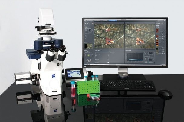
CellHesion 300 setup on Zeiss Axio Observer inverted microscope with intuitive software interface. Image Credits: Bruker Nano GmbH - JPK BioAFM
Applications
CellHesion data gallery
Bruker's BioAFMs enable biophysics and life science researchers to extend their examination in cell adhesion and mechanics, cell-cell and cell-surface interactions, mechanobiology, cell morphology, and cell dynamics. The company has gathered a gallery of images that establish some of these applications.
Key Features
Higher throughput
Improved productivity
Increased sample size and automated processes offer the statistical importance needed in pre-clinical and biomedical research.
Biomechanics
SmartMapping feature
Effortless and quick selection of several areas of interest over large sample regions. Flexible, user-defined 2D-shaped force maps.
Label-free measurements
Tissue biopsies
In near-physiological conditions, multiparametric, nanomechanical representation of living samples. Suitable for soft, highly topographic tissue samples and multi-layered cell structures.