The ImageXpress™ HCS.ai High-Content Screening System is an automated imaging platform designed for high-content analysis, capturing and evaluating cellular images in cell-based assays. This system facilitates the image-based assessment of various cellular characteristics by integrating automated microscopy with AI-enhanced image analysis in high-content screening processes.
The ImageXpress HCS.ai system works alongside MetaXpress® Acquire Software to facilitate automated image acquisition and connects with IN Carta® Image Analysis Software for AI-enhanced image analysis. Its adaptable structure accommodates various 2D and 3D cell-based assays and enables the configuration of the system to grow as the needs of the laboratory change.
Modular and intuitive for optimal versatility
The ImageXpress HCS.ai system boasts a flexible architecture along with numerous adjustable setup alternatives to cater to existing demands and accommodate on-site enhancements for future needs.
The recently improved MetaXpress® Acquire software serves as a user-friendly option suitable for individuals at any skill level, granting quick access to dependable and informative data in just a few minutes.
Higher quality images and superior data analysis for deeper insights
Obtain high-quality images with twice the signal-to-background ratio across a wide array of assay categories, achieve outstanding image clarity from 3D samples, and make new breakthroughs with the AI-driven IN Carta® Image Analysis Software.
Twice the acquisition speed for accelerated high-throughput workflows
Enhance laboratory efficiency by capturing more plates in less time and making it easier for newcomers to learn. A newly designed, automation-compatible framework allows users to effortlessly incorporate it into automated processes for quicker and more dependable outcomes.
Discover the new ImageXpress HCS.ai High-Content Screening System. Video Credit: Molecular Devices UK Ltd
What does the AI component of the ImageXpress HCS.ai system do?
The ImageXpress HCS.ai High-Content Screening System's AI feature aids image analysis by helping to locate and measure characteristics within cellular images. This technology is utilized in the analysis phase following acquisition to facilitate the handling of extensive image datasets and derive quantifiable metrics from cell-based experiments.
At a broader perspective, AI-assisted analysis may encompass:
- Automated identification of cellular features in acquired images
- Measurement of predefined image-based parameters
- Assistance for uniform analysis over extensive image datasets
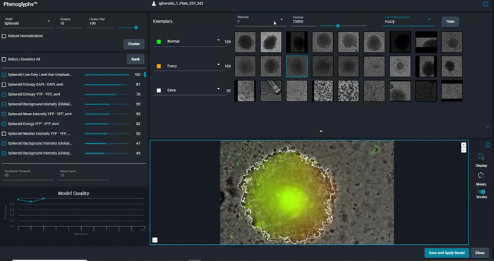
Image Credit: Molecular Devices UK Ltd
Where the ImageXpress HCS.ai High-Content Screening System fits in a typical experiment
The ImageXpress HCS.ai High-Content Screening System is used in the imaging and evaluation phases of a cell-based study. Once samples are arranged in line with the experimental guidelines, the system captures cellular images and assists in subsequent image evaluation. The outputs from this analysis can subsequently contribute to the larger experimental dataset.
At a high level, a standard workflow consists of:
- Preparing cell-based samples utilizing recognized experimental techniques
- Image acquisition through the ImageXpress HCS.ai system
- Analysis of images after acquisition with associated analysis software
- Creation of quantitative data derived from images for additional evaluation or integration
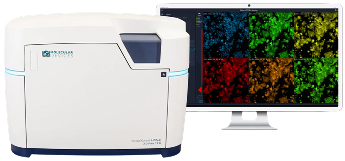
Image Credit: Molecular Devices UK Ltd
Features
Multiple imaging modes
The system provides brightfield label-free imaging, along with widefield and confocal fluorescent imaging.
Streamlined upgrade pathway
The modular design allows for simple integration of new features into the system, facilitating seamless upgrades as the requirements change over time.
High-intensity laser light source option
The use of laser illumination allows users to capture images more rapidly by using shorter exposure times and to conduct multiple experiments simultaneously with seven lasers and eight imaging channels.
MetaXpress® Acquire software
The MetaXpress® Acquire acquisition package simplifies intricate processes; new users can quickly begin using it, while experienced users can leverage its adaptability to develop enhanced protocols.
Automated water immersion objective technology option
Provides enhanced image resolution and sensitivity with a signal increase of up to four times, resulting in reduced exposure durations.
Exclusive AgileOptix™ spinning disk technology
Offers enhanced sensitivity through the use of specially designed optics, powerful laser lighting, and an sCMOS sensor. Swappable disk geometries allow for adaptability between rapid performance and high resolution.
Magnification changer option
Gain access to 12 different magnifications within a single configuration, featuring high-NA (Numerical Aperture) and ELWD (Extra-Long Working Distance) options.
AI-powered IN Carta® image analysis software
The software utilizes machine-learning techniques to enhance the precision and reliability of high-content image analysis, providing data insights that alternative technologies often overlook. Simplifies the process of image analysis through user-friendly guided workflows presented in a contemporary user interface.
Deeper insights for every lab
The ImageXpress HCS.ai High-Content Screening System with AI-powered insights
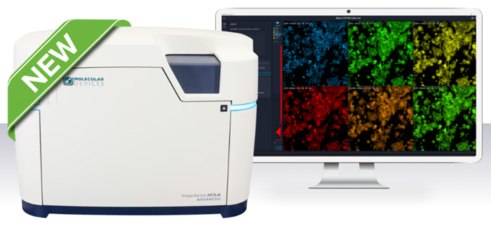
Image Credit: Molecular Devices UK Ltd
LED or laser illumination
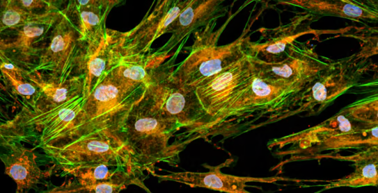
Cell painting image at 60X magnification captured under high-intensity laser illumination. Image Credit: Molecular Devices UK Ltd
LED light source
- Gentle illumination suitable for up to seven fluorescent channels
- DAPI, CFP, FITC, YFP, TITC, TxRed, Cy5
- Perfect for widefield, confocal, and conventional screening assays
Seven-laser high-intensity light source
- Intense excitation that supports up to eight distinct fluorescent channels
- DAPI, CFP, FITC, YFP, TRITC, TxRed, Cy5, Cy7
- Near-infrared illumination facilitates extensive spectral differentiation and a variety of fluorophore selections
- Enhance acquisition rates by markedly decreasing exposure durations, making it suitable for three-dimensional and highly multiplexed assays
AgileOptix spinning disk confocal
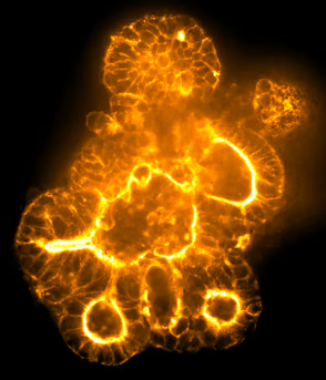
Organoid image acquired with a 60 µm pinhole confocal disk. Image Credit: Molecular Devices UK Ltd
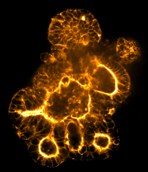
Organoid image acquired with a 50 µm pinhole deep tissue disk. Image Credit: Molecular Devices UK Ltd
The technology offers various confocal disk designs, encompassing both pinhole and slit arrangements for applications requiring high throughput.
- Standard disk (single): 60 µm pinhole disk, ideal for a broad spectrum of two-dimensional and three-dimensional applications
- Screening disk (dual): A combination of a 60 µm pinhole disk and a 50 µm slit disk, designed for enhanced screening speeds
- Deep tissue disk (dual): Consists of 50 µm pinhole disks featuring smaller pinholes and tighter spacing, tailored for increased signal detection or deeper sectioning within three-dimensional structures
Six objectives with water immersion
Six-position automated objective changer with optimal water immersion
The system can be equipped with up to six objectives simultaneously, offering magnification levels ranging from 2× to 60×, including high-numerical-aperture and extra-long-working-distance objectives, thereby minimizing the need for configuration adjustments.
The system can also accommodate up to two water-immersion objectives. These objectives provide signal enhancements of up to four times, assisting researchers in examining thicker and three-dimensional tissue samples with reduced exposure durations.
Magnification changer
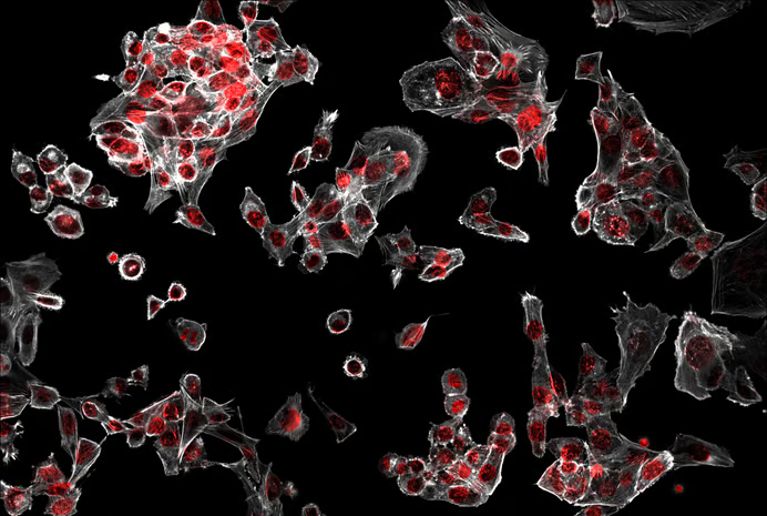
PC-12 cells captured at 40X magnification. Image Credit: Molecular Devices UK Ltd
Automated magnification changer
The ImageXpress HCS.ai system may be fitted with a magnification changer that enables the transition between 1X and 1.5X. This feature permits as many as 12 different magnifications within a single setup, eliminating the necessity to change objectives.
sCMOS sensor
sCMOS sensor with exceptional sensitivity and wide field of view
The >5 megapixel sCMOS sensor features a substantial sensor area of 224 mm2 and a dynamic range exceeding 3 log units with a 16-bit detector, achieving 95% peak quantum efficiency and enabling users to capture clearer images in less time.
MetaXpress Acquire High-Content Image Acquisition Software
Intuitive software for capturing images designed for various applications, fine-tuned for the ImageXpress HCS.ai platform
The revised MetaXpress® Acquire High-Content Image Acquisition Software simplifies intricate tasks. New users can quickly begin their journey thanks to the user-friendly, revamped interface and supportive workflows, while experienced users can leverage its adaptability to develop efficient procedures.
IN Carta Image Analysis Software
AI-driven analytics paired with an easy-to-use interface.
The advanced capabilities of IN Carta® image analysis software provide the tools users need to perform large-scale 2D, 3D, and 4D data analysis and deliver immediate insights without requiring complicated pre- or post-processing tasks.
Enhance the precision of image analysis workflows by leveraging the SINAP (Segmentation Is Not A Problem) deep-learning module. Employ machine learning to assist users and conduct intricate phenotypic analysis through an accessible Phenoglyphs module.
QuickID™ targeted image acquisition
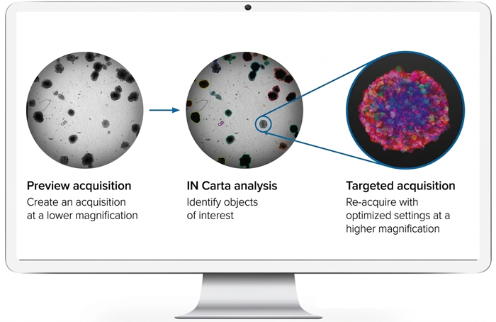
Train your system to recognize objects of interest with IN Carta deep-learning segmentation. Image Credit: Molecular Devices UK Ltd
AI-driven analytics paired with an easy-to-use interface
QuickID Targeted Acquisition operates by capturing a complete field of view of a sample at a low magnification. It identifies objects of interest or rare occurrences through rapid image analysis, then reacquires only these specific wells or locations at a higher magnification using a user-defined protocol.
This protocol may encompass the necessary wavelengths, z-stacks, and time points. As a result, this approach facilitates quicker image acquisition, reduces the number of images that need to be analyzed, and requires less data storage space, while also ensuring that the object of interest is consistently centered.
Scalable by design. Powerful by choice
The ImageXpress® HCS.ai system features a modular design that scales with research. Whether users begin with widefield imaging or analyze confocal and deep tissue, this platform adapts to their requirements, therefore eliminating the need for a complete system overhaul.
A wide array of configuration possibilities and user-friendly acquisition software simplify the processes of upgrading, adapting, and expanding, ensuring the investment remains relevant and facilitating faster discoveries.
The majority of upgrades can be implemented directly on-site by skilled technicians, reducing downtime and enhancing flexibility.
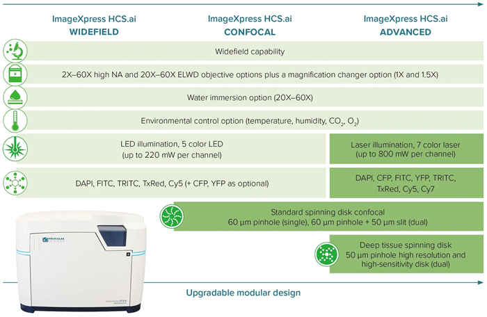
Image Credit: Molecular Devices UK Ltd
See the science. Capture the complexity
The ImageXpress HCS.ai system empowers individuals to conduct a range of assays on more physiologically relevant cell models, facilitating deeper understanding and more significant findings.
Obtain remarkable, high-resolution images from prolonged live-cell studies, 3D structures, and multiplexed phenotypic assays using the user-friendly MetaXpress® Acquire software. Enhance the analysis process with the AI-driven IN Carta® Image Analysis Software, designed to unravel even the most intricate biological inquiries swiftly and accurately.
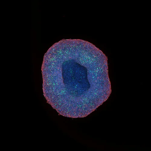
HepG2 spheroid acquired at 10X magnification. Image Credit: Molecular Devices UK Ltd
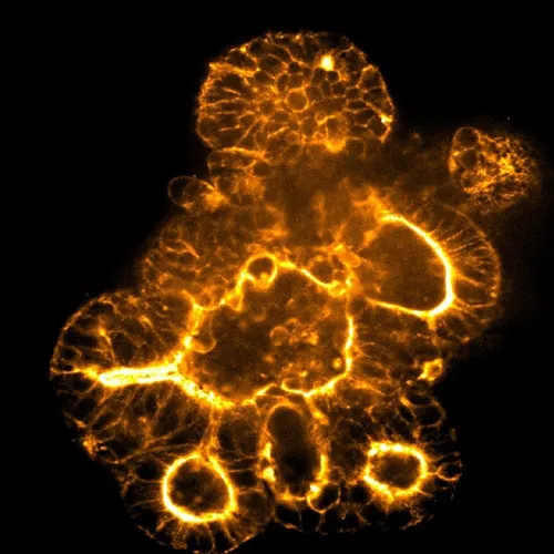
Organoid image acquired at 20X magnification. Image Credit: Molecular Devices UK Ltd
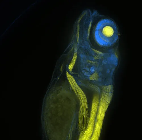
Zebrafish embryo acquired at 10X magnification. Image Credit: Molecular Devices UK Ltd
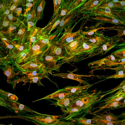
Cell painting assay at 60X magnification. Image Credit: Molecular Devices UK Ltd
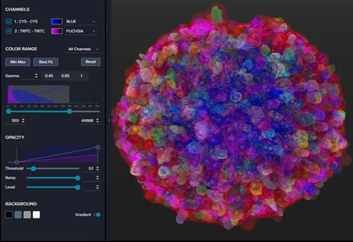
Spheroid mask in 3D viewer in IN Carta Image Analysis Software. Image Credit: Molecular Devices UK Ltd
Scalable walkaway automation: Flexible solutions for high-content screening with the ImageXpress HCS.ai system
For laboratories starting their automation journey, Molecular Devices provides adaptable and expandable solutions that ease the transition toward automated high-content screening workflows. Starter automation options, like walkaway automation for the ImageXpress™ HCS.ai High-content Screening System, enhance plate management and combine sophisticated imaging with AI-driven analysis.
With this setup, users can image more plates in less time; they can process 40 microtiter plates (96-well format) within just two hours, or 80 plates in four hours, all without needing hands-on intervention.
This method increases throughput, guarantees high-quality data, and reduces the need for manual tasks. As research progresses, these modular systems can expand from simple plate handling to fully integrated, customized workcells, providing consistent, reproducible outcomes with maximized efficiency.
There are significant benefits to scalable laboratory automation:
- Walkaway efficiency: Handle hundreds of plates without supervision, freeing up important time for laboratory personnel
- Increased throughput and enhanced reproducibility: Automated imaging guarantees uniform and dependable outcomes
- Effortless integration: Works with the chosen software and laboratory configuration seamlessly
- Future-ready: Facilitate simple upgrades as new features become available
- Modular and scalable: Expand the system as research grows
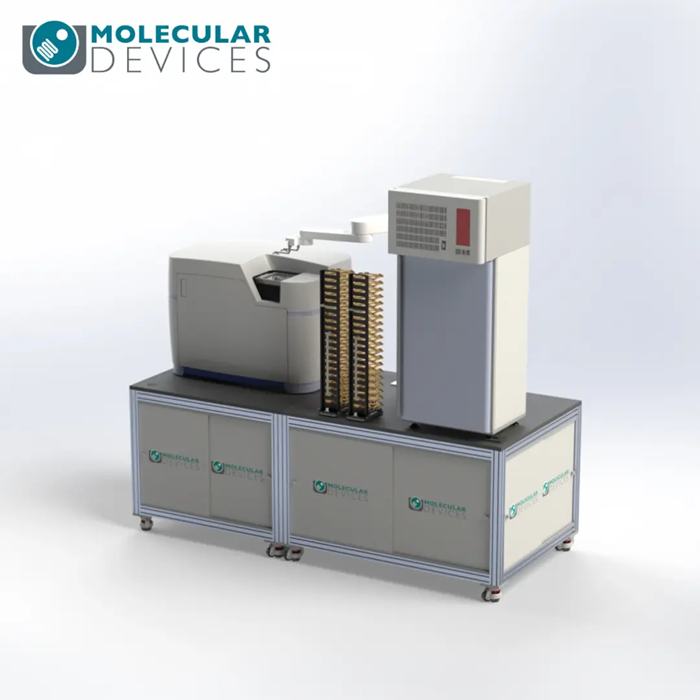
Image Credit: Molecular Devices UK Ltd
Applications
Applications of the ImageXpress HCS.ai High-Content Screening System
3D cell imaging and analysis
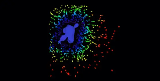
Image Credit: Molecular Devices UK Ltd
Three-dimensional (3D) cell models are relevant to physiological conditions and provide a closer approximation of tissue microenvironments, interactions between cells, and the biological processes that take place in a living organism.
Currently, users can produce more reliable data by using the ImageXpress system with the integrated 3D Analysis Module in MetaXpress® software. This unified platform will enable users to address challenges in 3D data acquisition and analysis without sacrificing efficiency or data quality, instilling confidence in their findings.
COVID-19 and infectious disease research
Molecular Devices has covered typical uses in the study of infectious diseases, such as the development of cell lines, assessment of binding affinity, viral neutralization, evaluation of viral titer, and additional topics. Their emphasis has been on comprehensively understanding the SARS-CoV-2 virus to facilitate the creation of potential treatments for COVID-19, which include vaccines, therapeutic agents, and diagnostic tools.
Cell painting
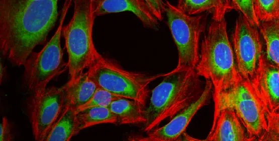
Image Credit: Molecular Devices UK Ltd
Cell painting is a complex imaging assay that employs fluorescent dyes to mark various cellular components for extensive phenotypic analysis.
High-content imaging facilitates the automated acquisition and examination of cell painting data, aiding in the extraction of features across organelles to identify subtle phenotypic alterations induced by treatment.
Cells in extracellular matrices
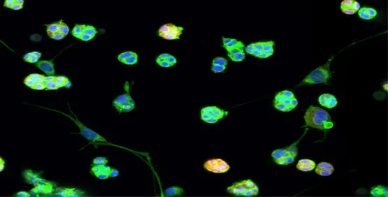
Image Credit: Molecular Devices UK Ltd
One widely used technique to cultivate cells in a three-dimensional environment leverages hydrogels based on extracellular matrix, for instance, Matrigel. Cells are cultivated within an extracellular matrix (ECM) to simulate an in vivo setting.
Distinctions between Matrigel and two-dimensional cell cultures are clearly observable in features such as cell shape, cell orientation, and gene expression patterns. Furthermore, hydrogels facilitate investigations into cell migration and the formation of three-dimensional structures, including the development of endothelial cell tubes in studies of angiogenesis.
Disease modeling
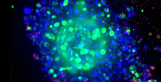
Image Credit: Molecular Devices UK Ltd
Disease model systems vary significantly in terms of complexity and scale, spanning from basic two-dimensional cell cultures to intricate model organisms. Although model organisms provide a living context for investigation, they can often be expensive and might not accurately reflect human biology.
Conversely, traditional two-dimensional cell culture systems, which have been used for many years, struggle to replicate the complex three-dimensional architecture and cellular interactions inherent in living tissues. Consequently, three-dimensional cell cultures have appeared as a promising alternative model system for disease modeling.
Drug discovery and development
High-content imaging is used in drug discovery and development to evaluate how cells respond to various compounds using multiple biological indicators.
By collecting quantitative, image-based information on a large scale, researchers are able to assess phenotypic impacts, alterations related to mechanisms, and cellular toxicity throughout preclinical research processes.
Live-cell imaging
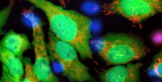
Image Credit: Molecular Devices UK Ltd
Live-cell imaging is employed to monitor cellular processes over time while maintaining controlled experimental conditions.
This method enables researchers to observe evolving occurrences, including alterations in cell structure, movement, or actions throughout an experiment. Live-cell imaging generally requires controlling environmental conditions and capturing images over time.
Neurite outgrowth/tracing
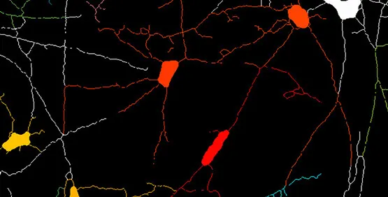
Image Credit: Molecular Devices UK Ltd
Neurite outgrowth assays serve to assess the elongation and division of neuronal extensions either during the developmental phase or in reaction to specific experimental circumstances.
High-content imaging facilitates the automation of neurite tracing and the quantitative evaluation of factors such as neurite length, branching complexity, and neuronal morphology across various cell populations.
Oncology
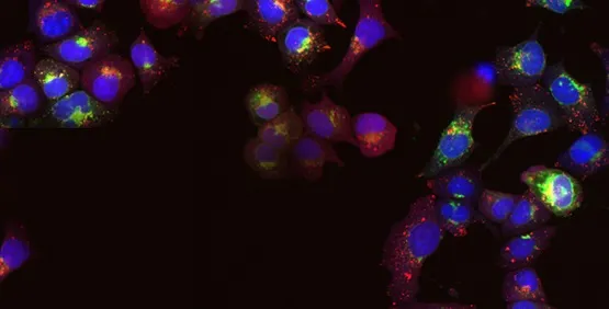
Image Credit: Molecular Devices UK Ltd
In oncology research, high-content imaging is utilized to investigate the behavior, structure, and reaction of cancer cells to various experimental conditions at the cellular level.
This method facilitates the examination of diverse cell populations, allowing for evaluation based on images to identify variations in phenotypes, alterations in the cell cycle, or effects resulting from treatments in cancer models.
Organ-on-a-chip technology
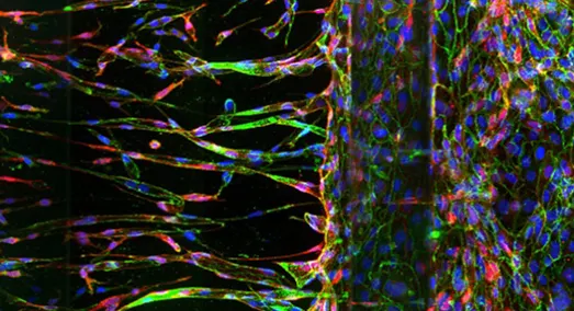
Image Credit: Molecular Devices UK Ltd
Organ-on-a-chip (OoC) technology uses microfabrication methods to develop small-scale representations of biological organs, such as the lung, heart, or intestine, within a chip-sized apparatus.
These miniaturized devices consist of living cells cultivated on a microscale platform, imitating the architecture and function of the associated organ. The cells are usually positioned to replicate the organ's natural three-dimensional form and supplied with fluids, such as blood or air, to simulate its physiological conditions.
Organoids
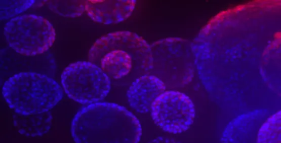
Image Credit: Molecular Devices UK Ltd
Organoids are three-dimensional (3D) multicellular microtissues intended to accurately replicate the intricate structure and function of human organs. Typically, organoids are composed of a co-culture of cells that exhibit a high degree of self-assembly, enabling a more precise representation of complex in vivo cellular responses and interactions than conventional two-dimensional (2D) cell cultures.
Spheroids
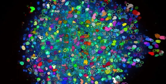
Image Credit: Molecular Devices UK Ltd
Spheroids are three-dimensional structures composed of multiple cells that replicate the responses and interactions of cells in a living organism. These structures can be produced with high consistency and can be adapted for extensive screening processes.
In contrast to adherent cells cultivated in flat layers, it is thought that growing cells in 3D conditions more accurately represents the actual environment of cancer cells. Gathering data from these larger formations requires capturing images at various depths (z-planes) within the spheroid and examining them in three dimensions, or compressing them into a unified two-dimensional stack for analysis.
Stem cell research
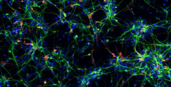
Image Credit: Molecular Devices UK Ltd
Pluripotent stem cells are suitable for developmental biology research and can be differentiated to generate organ-specific cells, which can be used in live- or fixed-cell-based assays on slides or in multi-well plates. The ImageXpress™ system is beneficial throughout the entire process of stem cell research, from monitoring differentiation to ensuring quality control and assessing the functionality of particular cell types.
T-cells and 3D cell models for immunotherapy
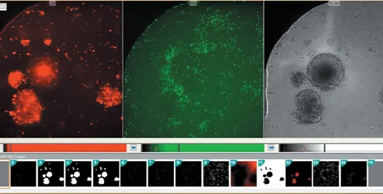
Image Credit: Molecular Devices UK Ltd
Harnessing T-cells alongside organoids and spheroids for cancer therapy investigation. Immunotherapy, which utilizes the body’s immune mechanisms to precisely attack cancer cells, is increasingly being recognized as a complementary approach to cancer treatment. This category of therapies encompasses CAR T-cells (Chimeric Antigen Receptor-engineered T-cells), tumor-infiltrating lymphocytes (TILs), and various genetically altered T-cells.
Enhancing the recruitment of T-cells and their ability to destroy cancer cells is vital for effective cancer immunotherapy. In preliminary scientific studies, three-dimensional cell models offer benefits over two-dimensional monolayer cultures, as they more accurately simulate the immunosuppressive tumor microenvironment (TME). For instance, organoids derived from patients (PDOs) outperform conventional 2D monolayer cultures by more faithfully recapitulating the physical and chemical signals of the TME.
Toxicology
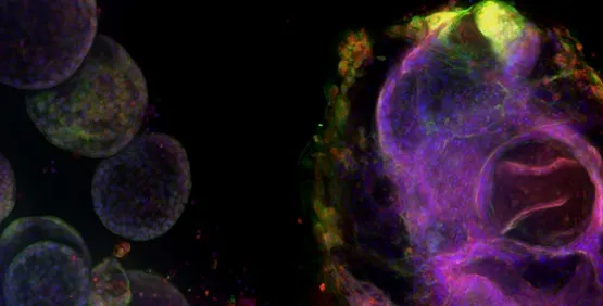
Image Credit: Molecular Devices UK Ltd
Toxicology examines the harmful impacts of both natural and man-made chemicals on living organisms. Today, it is an increasingly significant issue as everyone comes into contact with a greater number of chemicals present in both their surroundings and the items they use.
Specifications and options
Source: Molecular Devices UK Ltd
|
| . |
. |
| Focus |
- High-speed hardware autofocus with multi-surface detection
- Integrated image autofocus option
|
| Camera |
- >5 megapixels sCMOS sensor with large sensor size (224mm2)
- >3 log dynamic range with 16bit detector, 95% peak quantum efficiency
|
| Stage |
Linear encoded voice coil driven X, Y, and Z stages with better than 25 nm resolution |
| Filters |
- Four-position software selectable excitation and dichroic filter wheel
- 10-position software selectable emission filter wheel
|
| Transmitted light |
High-contrast imaging where unstained cells are easily viewed or separated from background |
| Objectives |
- Six-position automated objective changer
- High NA air objectives: 2X–60X (up to 0.85 NA)
- Extra long working distance objectives 20X–60X (up to 0.7 NA)
- Water immersion (20x, 40x, 60x)
|
| Sample compatibility |
Microplates (1–1536 wells), round or flat bottom, low to high profile, slide holder |
| Magnification changer (optional) |
- 1X (standard) and 1.5X modifies objective capability: to provide 2X–90X magnification
- Automated switching with software control
|
| Water immersion (optional) |
- 20X, 40X, 60X objectives available (up to 1.2 NA)
- Increase signal up to four times for brighter intensity at lower exposure times
- Increase in penetration depths dependent on sample
- Improve z-resolution and decrease optical aberrations
- Auto water replenishment enables screening or imaging across a plate
|
| Environmental Control (optional) |
- Multi-day, live cell time-lapse imaging
- Controls for appropriate atmospheric conditions
- Temperature: heating up to 40 °C
- Integrated gas mixer: CO2 (0–15%), O2 (1–21%)
- Controls humidity and minimizes evaporation (0.5 µL/well/hour for 96- or 384-well formats)
- Robot-friendly loading design
|
| Autofocus |
Hardware and Image autofocus |
| Plates supported |
Up to 1536-well plates |
| Temperature control |
✔ |
| Maximum Illumination Channels |
Seven |
| Robotics/Automation-compatible |
✔ |
Source: Molecular Devices UK Ltd
| Model |
ImageXpress.HCS.ai Widefield |
ImageXpress.HCS.ai Confocal |
ImageXpress.HCS.ai Advanced |
| Widefield capability |
Yes |
Yes |
Yes |
| Water immersion option (20X-60X) |
Yes |
Yes |
Yes |
| Magnification changer option (1X and 1.5X) |
Yes |
Yes |
Yes |
| Environmental control option |
Yes |
Yes |
Yes |
| LED illumination |
Yes |
Yes |
|
| Standard spinning disk confocal (60 µm pinhole (single), 60 µm pinhole + 50 µm slit (dual)) |
|
Yes |
Yes |
| Deep tissue spinning disk 50 µm pinhole Deep Tissue disk (dual) |
|
|
Yes |
| Laser illumination |
|
|
Yes |