Bruker’s Papilio5D light-sheet microscope is built to give researchers high-quality imaging with exceptional depth penetration - even in dense samples like organoids or zebrafish larvae.
The system combines dual-view imaging with modular illumination beams, maximizing photon efficiency and improving image clarity. With dual-sided detection, it captures twice the photon signal compared to single-objective setups, offering a more complete 3D view of your samples.
Users can select between Gaussian or Bessel beam illumination, each with a beam width under one micrometer, depending on their application needs. The flexible sample mounting system supports complex experimental setups, including multiplexing and pharmacology studies.
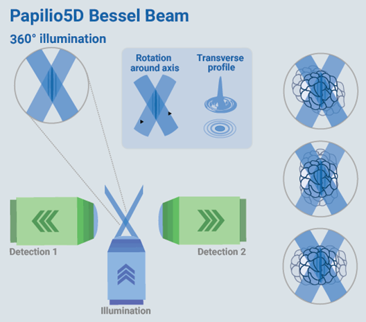
Image Credit: Bruker Nano Surfaces and Metrology
Only Papilio5D offers:
- Quick dual-view acquisition and fusion for high-resolution images
- Ideal light-sheet imaging of many samples under different experimental conditions at the same time
- Dual-sided detection and modular illumination beams for the highest photon collection efficiency
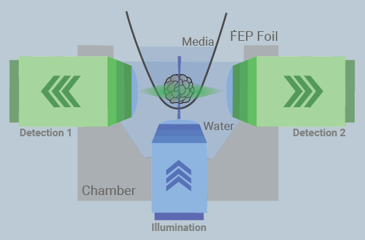
Papilio5D uses horizontal dual-view detection and a choice of Bessel or Gaussian illumination. This setup optimizes depth penetration and provides a large and evenly illuminated field of view with four different optical magnifications. Image Credit: Bruker Nano Surfaces and Metrology
Customizable solutions for long-term imaging
Highest image quality
Papilio5D features a horizontal imaging geometry with dual-view detection and perpendicular light-sheet illumination, making it ideal for imaging even the most delicate samples with maximum photon efficiency.
High-NA water-dipping lenses create an ultra-thin light sheet for precise imaging, while both area and line detection modes allow users to tailor data acquisition to specific sample types. This setup delivers a high signal-to-noise ratio, excellent Z resolution, and strong depth penetration.
Papilio5D can also support Nyquist sampling, enabling high-fidelity image reconstruction and advanced deconvolution for clearer, more accurate results.
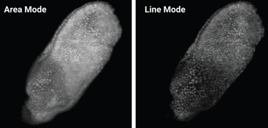
Comparison of area vs. line mode. Area mode images quickly and efficiently, while in line mode, the camera uses a rolling shutter and improves contrast in thick samples. Image Credit: Bruker Nano Surfaces and Metrology
Simple and intuitive sample mounting
Gentle, flexible sample mounting is essential for running multi-sample and multi-condition experiments, and Papilio5D is built with that in mind.
Its spacious sample holder accommodates up to three dishes, each with two separate chambers, allowing researchers to test six experimental conditions simultaneously.
To support long-term imaging, from hours to days, the system includes full environmental control, maintaining sample health and stability. Users can regulate temperature (20–37 °C), gas concentrations (CO2 and O2), and humidity, ensuring optimal conditions for live samples throughout extended experiments.
Optimized beam patterns for different applications
With Papilio5D, scientists can tailor light-sheet imaging to suit their specific experimental needs.
A Gaussian beam is ideal for small, light-sensitive samples - such as those used in early mouse embryology - offering gentle illumination and high precision. For imaging more optically dense samples, the Bessel beam provides 360° multi-directional illumination, ensuring deeper penetration and more uniform coverage.
Researchers can also choose between two acquisition modes to match their imaging goals. In area mode, the camera captures the full field of view in a single shot, making it fast and efficient for many applications.
In line mode, the system uses the camera’s rolling shutter in a confocal slit-like configuration, synchronized with the scanned light-sheet. This setup rejects out-of-focus and scattered light, significantly improving contrast in thick or scattering samples - especially useful when working with complex structures like organoids.
Papilio5D specifications
Source: Bruker Nano Surfaces and Metrology
| Dual Sided Detection with 2x Objectives |
Tube
Lens |
Magnification |
Pixel Size [nm]
Orca Flash |
FOV [μm]
Orca Flash |
Pixel Size [nm]
Orca Fire |
FOV [μm]
Orca Fire |
| Detection 2x Nikon CFI APO LWD 25x 1.10 NA |
100 |
12.5x |
520 |
1065 |
370 |
876 |
| 200 |
25x |
260 |
532 |
184 |
436 |
| 300 |
37.5x |
173 |
353 |
123 |
290 |
| 400 |
50x |
130 |
266 |
92* |
218 |
| Detection 2x Nikon CFI APO LWD 16x 0.8 NA |
100 |
8x |
813 |
1664 |
575 |
1362 |
| 200 |
16x |
406 |
832 |
288 |
681 |
| 300 |
24x |
271 |
555 |
192 |
454 |
| 400 |
32x |
203 |
416 |
144 |
340 |
Illumination
Modes |
Gaussian
Beam |
Bessel
Beam |
|
|
|
|
Beam Waist
Thickness [μm] |
1 to 6 |
1 (main lobe) |
|
|
|
|
| Beam Length [μm] |
20 @ 1 μm waist |
350 |
|
|
|
|
| Pivot Scan |
YES |
YES |
|
|
|
|
| Confocal Line Mode |
YES |
YES |
|
|
|
|
| Acquisition Options |
- Gaussian beam
- Gaussian with line mode
- Gaussian with line mode and pivot scan
|
- Bessel beam
- Bessel beam with line mode
- Bessel beam with line mode and pivot scan
|
|
|
|
|
| Illumination Optics |
20x 0.6 NA |
|
|
|
|
|
| Detection Optics |
Dual 25x 1.1 NA/16x 0.8 NA |
|
|
|
|
|
Multi-Sample/Multi-
Condition Experiments |
6 separate compartments |
|
|
|
|
|
| Magnification Changer |
4 discrete steps |
|
|
|
|
|
| Data Processing |
Content based dual view registration and fusion |
|
|
|
|
|
| Photomanipulation |
CW or pulsed, VIS to NIR |
|
|
|
|
|
*Nyquist sampling