With its high-resolution imaging and potent analytic capabilities, the ImageXpress® Pico Automated Cell Imaging System is more than just a digital microscope.
The automated imager often offers a wide range of predefined procedures for cell-based assays, whether that be for fluorescence imaging or brightfield assays. This shortens the learning curve and enables users to launch investigations quickly.
With features like Digital Confocal 2D on-the-fly deconvolution, Live Preview, Autofocus, multi-wavelength cell scoring, and an optional workflow for IN Carta® Image Analysis Software, the ImageXpress Pico gives users the opportunity to advance their discoveries in a small and relatively inexpensive imager.
Get started quickly
CellReporterXpress® Image Acquisition and Analysis Software, which is icon-driven and simple to use, can expedite digital microscopy throughout the laboratory. Additionally, users can begin capturing and examining images with minimal training.
Do more than cell counting
Users can expand assays with more than 25 preconfigured templates improved for several cell-based experiments such as apoptosis, 3D cell models, mitochondrial evaluation, multiwavelength cell scoring, live cell/timelapse, and neurite tracing.
Automate imaging affordably
This system also removes the hassle of running samples to the core lab. The system’s lab-friendly price gives researchers the convenience of being able to automate imaging and analysis on their lab bench. With options like Digital Confocal, environmental control and z-stack acquisition, the system can be ordered to fit the users' research.
ImageXpress Pico Automated Imaging System Virtual Tour.Video Credit: Molecular Devices UK Ltd
Features
Multiple imaging modes
With objectives ranging from 4× to 63×, the ImageXpress Pico system can be utilized for colorimetric, fluorescence, brightfield, or digital confocal 2D on-the-fly deconvolution imaging modes.
Preconfigured analysis protocols
More than 25 preconfigured analysis protocols are available, ranging from simple cell counting to sophisticated neurite tracing analysis. By simply clicking on a few cells that satisfy a certain set of requirements, analytical parameters can be adjusted using tools like the click-to-find tool.
Plate-to-individual cell view
Data can be envisioned at several levels, from plate overview to individual cells. Users can access a wide variety of data visualization tools from the cellular to the plate level, enabling them to extract a wealth of knowledge from their images and assays.
Z-stack acquisition
Generate sharper images for more accurate segmentation using z-stack acquisition. Users can also acquire a series of images at different focal points to capture more detail than is possible with a single slice. Users can include all slices or select which slices to include in the final projection.
Quickly and easily identify regions of interest
By enabling users to interactively alter the focus with a virtual joystick while panning over the sample, Live Preview streamlines the identification of regions of interest and saves time and effort.
Environmental control
The onboard environmental system, which has options for controlling CO2, humidity, and O2, can be utilized to execute multi-day, live cell assays and time-lapse experiments. The software is also enhanced to prevent z-drift and provides real-time environmental monitoring, ensuring superior assay conditions.
Experience the powerful combination of the ImageXpress Pico and CellReporterXpress
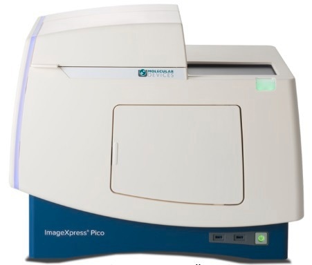
ImageXpress Pico Automated Cell Imaging System. Image Credit: Molecular Devices UK Ltd
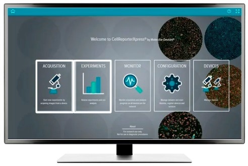
CellReporterXpress Image Acquisition and Analysis Software. Image Credit: Molecular Devices UK Ltd
CellReporterXpress software for ImageXpress Pico
Easy-to-learn software optimized for automated digital microscopy
A simple and intuitive interface with features like Digital Confocal 2D real-time deconvolution, Autofocus, and Live Preview for sophisticated regions of interest identification is provided to allow users to perform quantitative analysis on images obtained from automated microscopy.
The software enables distributed analysis of images for increased throughput and is ideal for scaling digital microscopy imaging with slides or microplates. An icon-driven, linear workflow with a range of predefined protocols provides a streamlined user experience.
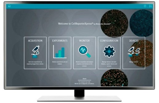
Image Credit: Molecular Devices UK Ltd
IN Carta® Image Analysis Software
Go from assay to insights quickly and reliably
Workflows for phenotypic profiling and image analysis are made more accessible by robust analytics combined with a simple user interface. Advanced capabilities allow users to analyze data in 2D, 3D, and 4D at scale and provide real-time information without the need for laborious pre- or post-processing procedures.
By employing the SINAP deep-learning module, users can improve the precision of their image analysis workflows and also see that segmentation is not a problem. Machine learning helps users perform complex phenotypic analysis within a user-friendly Phenoglyphs module.
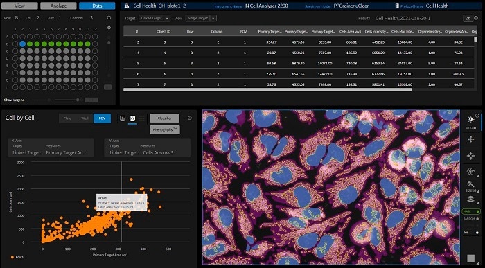
Image Credit: Molecular Devices UK Ltd
Tailored laboratory automation solutions with robotics, incubators, and software
Modern researchers want simplified remote access and enhanced laboratory automation as the research environment constantly changes. When the ImageXpress® Pico Automated Cell Imaging System is combined with the SCILA incubator from Inheco and the S-LABTM plate handler from PAA, high productivity, reduced costs, and consistent performance are all possible.
Automated ImageXpress Pico workflow in action. Video Credit: Molecular Devices UK Ltd