This article and associated images are based on a poster originally authored by Isabel Gameiro-Ros, Denise R B de Pilger, Ming Jiang, Ok-Ryul Song, Scott Warchal, Christophe Queval, Adrien Pasquier, Joseph Tuersley, Harry Saville, John Hellicar, Sara Tamagno and Michael Howell and presented at ELRIG Drug Discovery 2026 in affiliation with The Francis Crick Institute.
This poster is being hosted on this website in its raw form, without modifications. It has not undergone peer review but has been reviewed to meet AZoNetwork's editorial quality standards. The information contained is for informational purposes only and should not be considered validated by independent peer assessment.

Overview
Human induced pluripotent stem cells (iPSCs), embryonic stem cells (ESCs), and 3D organoids are becoming foundational models in drug discovery. They provide authentic, experimentally accessible human cells and improve the translational relevance of target and compound discovery. However, using them at scale remains difficult. Maintaining these cultures needs considerable technical skill and staff commitment, an estimated 10-20,000 hours per year across Crick stem cell researchers, and complex manipulations such as differentiations can demand up to 30 days of continuous activity. Manual handling also adds batch-to-batch and user-related variability that carries through into downstream assays.
To overcome these limitations, we developed integrated automated platforms that connect robust stem cell culture with genetic engineering, complex cell-based assays, and high-throughput screening. Here we show how automation can:
- Maintain multiple ES/iPSC lines long-term with minimal manual intervention
- Support CRISPR engineering and functional screening
- Enable multiplexed and continuous high-throughput screening on stem cell-based models
What an automated and integrated workflow enables
- Automated ES/iPSC culture: Single-cell and clump passaging, long-term, standardized and monitored.
- Robust QC of stem cells: Ensure ES/iPSCs maintain quality standards in automated culture.
- Genetic engineering: CRISPR-based cell line engineering.
- High-throughput screening: CRISPR KO/overexpression, small molecule and siRNA screens.
- Downstream models: Stem cell differentiation and organoid generation.
Infrastructure: Automated platforms
1. Automated stem cell culture
The platform consists of multiple devices that interact with each other and communicate via Momentum scheduling software. Robotic handling, automated liquid handling, incubation, and imaging enable long-term, standardized maintenance of multiple human ES/iPSC lines with minimal manual intervention. Our workflows support both single-cell and clump passaging, with automated monitoring of culture quality and cell state through live imaging.
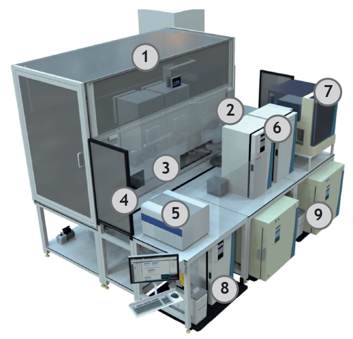
Fig 1. Automated stem cell platform. 1) BSL2 cabinet; 2) Spinnaker; 3) Tecan Fluent; 4) Washer and Lidpark; 5) Cytation 1 imager; 6) Incubators (small); 7) Tip/Plate hotel; 8) Fridge; 9) Incubators (large). Image Credit: Isabel Gameiro-Ros et al., in partnership with ELRIG (UK) Ltd.
2. Supporting automated infrastructure
- Liquid handling: Our portfolio (Echo, Combi nL, Myra, Biomek FX, Multiflo and Viaflo) lets us run complex cell-based and other assays in a range of plate formats (24- to 384-well) and volumes (nL to mL).
- Automated microscopes: Our imaging resources span from low-resolution live-cell imaging over several days to spinning-disk confocal microscopes (e.g., Opera Phenix Plus) coupled to robotic systems, capable of continuous and autonomous image acquisition and analysis.
- Automated analysis pipelines: Quantification of stem cell properties and cell line-specific characteristics of stem cell-based models.
- Database reference: Benchmark data mapped to internally established and standardized stem cell quality criteria.

Fig 2. Example of one of our automated microscopes and stem cell models generated in collaboration with Crick’s research groups. Image Credit: Isabel Gameiro-Ros et al., in partnership with ELRIG (UK) Ltd.
3. Stem cell-based 3D model systems
Our automated platforms are not confined to adherent cells. We also help research groups use them for 3D model systems, where they can support the maintenance, monitoring, and experimental manipulation of organoids, gastruloids, and cancer spheroids.
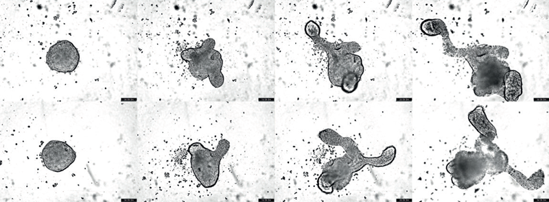
Fig 3. Brightfield images of trunkoids (days 0-3). In collaboration with Tiago Rito (Briscoe lab). Image Credit: Isabel Gameiro-Ros et al., in partnership with ELRIG (UK) Ltd.
Results: Quality and reproducibility of automated stem cell cultures
1. Pluripotency and genomic stability
Across 16+ passages, automated ES and iPSC cultures maintained genomic stability, > 95% expression of pluripotency markers, and robust trilineage differentiation capacity. STR profiling and mycoplasma testing confirmed the identity of all cell lines and the absence of contamination, respectively.
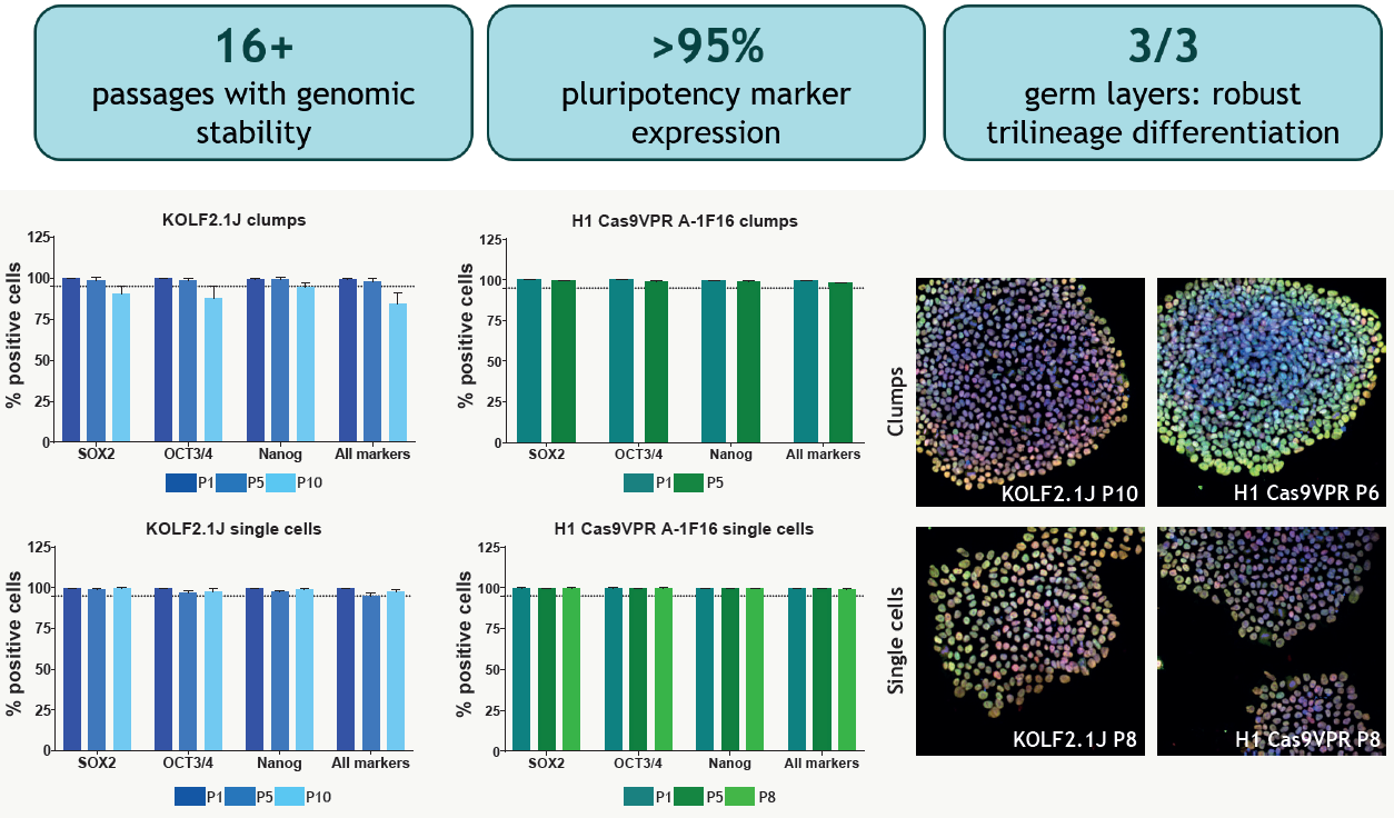
Fig 3. We tested the ability of the system to maintain human ES and iPSC cell lines in 12-well plates for a minimum of 6 and 8 passages, for clumps (panel A) and single cells (panel B) passaging (3-5 weeks). We observed reproducible cellular morphology over time in cultures maintained by the automation platform, consistent with standard PSC morphology. We set a threshold of > 95% of the population expressing combinations of three markers of undifferentiated state as an indicator of the quality of the cell culture. Image Credit: Isabel Gameiro-Ros et al., in partnership with ELRIG (UK) Ltd.
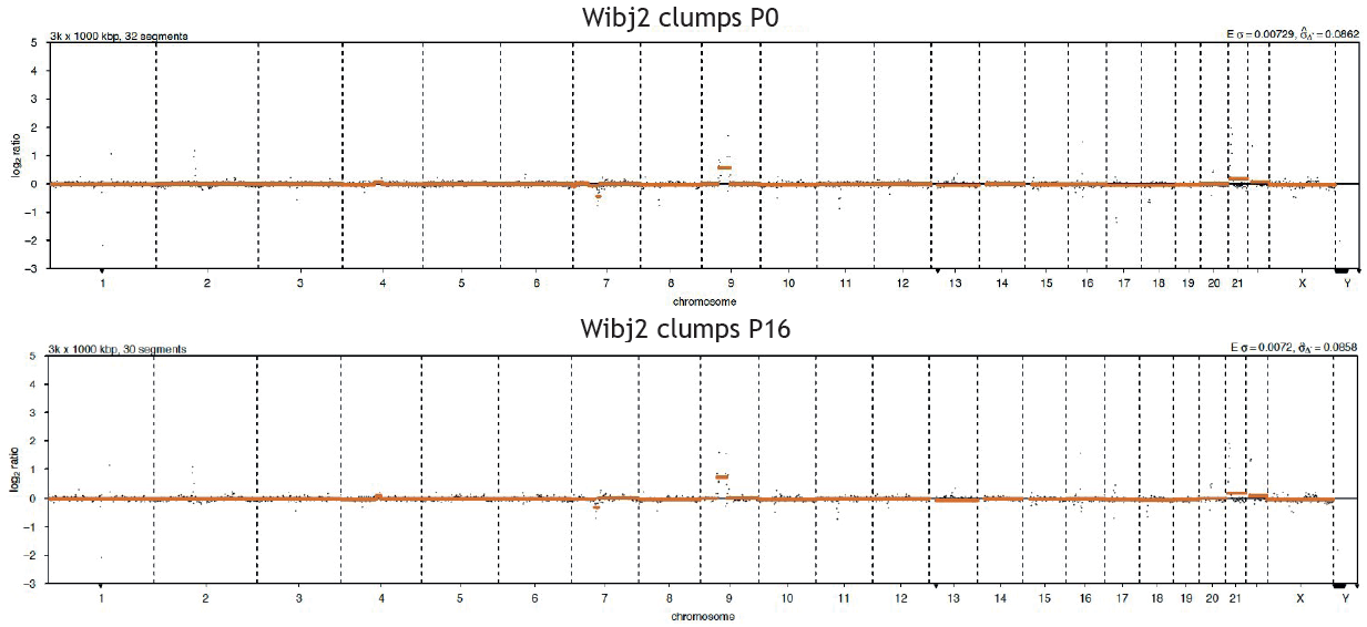
Fig 4. ES/iPSC samples were sent for Low-Pass Whole-Genome sequencing, showing no changes in the number or in the chromosomal structure over several passages (iPSCS shown, 16 passages, > 9 weeks). Cells maintained in our automated platform retained their healthy colony morphology, undifferentiated state and genomic stability over several passages. Image Credit: Low-Pass Whole-Genome sequency performed by the Genomics Science Technology Platform
2. Differentiation capability
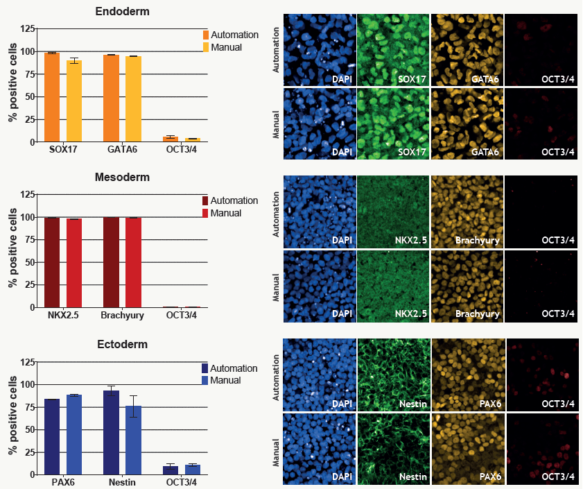
Fig 5. We used the STEMdiff™ Trilineage Differentiation Kit to compare the differentiation of Wibj2 iPSCs cells using the automation system and the manual method, obtaining similar results for both approaches: All three germ layers presented an elevated expression of specific markers for early differentiation and low expression of the undifferentiated marker OCT3/4. Image Credit: Isabel Gameiro-Ros et al., in partnership with ELRIG (UK) Ltd.
Conclusions
Our automated stem cell platform turns labor-intensive stem cell and complex cell-based workflows into a scalable resource for discovery research as well as drug and target discovery.
Automated cultures maintain genomic stability, pluripotency, and trilineage capacity over 16+ passages while cutting hands-on time by up to 85%.
Over 85% less hands-on researcher time
8 x 12-well plates, 4 cell lines, passaging every 4 days for 9 weeks
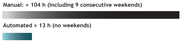
Image Credit: Isabel Gameiro-Ros et al., in partnership with ELRIG (UK) Ltd.
Our integrative approach, coupling automated stem cell culture with automated data analysis and quantification, allows us to streamline and positively contribute to the growing field of stem cell research.
Acknowledgments
We thank the Wolfson Foundation for funding the automated stem cell culture platform.
We also thank the Stem Cell and Automation Platform (SCOP) and the Genomics Science Technology Platform at the Francis Crick Institute for their collaboration on this work.
About The Francis Crick Institute
The Francis Crick Institute is dedicated to understanding the fundamental biology underlying health and disease. Our work is helping to understand why disease develops and to translate discoveries into new ways to prevent, diagnose and treat illnesses such as cancer, heart disease, stroke, infections and neurodegenerative diseases.
An independent organization, our founding partners are the Medical Research Council (MRC), Cancer Research UK, Wellcome, UCL, Imperial College London and King's College London.
The Crick was formed in 2015, and in 2016 we moved into our brand new state-of-the-art building in central London which brings together 1500 scientists and support staff working collaboratively across disciplines, making it the biggest biomedical research facility under a single roof in Europe.
About ELRIG (UK) Ltd.
The European Laboratory Research & Innovation Group (ELRIG) is a leading European not-for-profit organization that exists to provide outstanding scientific content to the life science community. The foundation of the organization is based on the use and application of automation, robotics and instrumentation in life science laboratories, but over time, we have evolved to respond to the needs of biopharma by developing scientific programs that focus on cutting-edge research areas that have the potential to revolutionize drug discovery.
Comprised of a global community of over 12,000 life science professionals, participating in our events, whether it be at one of our scientific conferences or one of our networking meetings, will enable any of our community to exchange information, within disciplines and across academic and biopharmaceutical organizations, on an open access basis, as all our events are free-of-charge to attend!
Our values
Our values are to always ensure the highest quality of content and that content will be made readily accessible to all, and that we will always be an inclusive organization, serving a diverse scientific network. In addition, ELRIG will always be a volunteer led organization, run by and for the life sciences community, on a not-for-profit basis.
Our purpose
ELRIG is a company whose purpose is to bring the life science and drug discovery communities together to learn, share, connect, innovate and collaborate, on an open access basis. We achieve this through the provision of world class conferences, networking events, webinars and digital content.
Sponsored Content Policy: News-Medical.net publishes articles and related content that may be derived from sources where we have existing commercial relationships, provided such content adds value to the core editorial ethos of News-Medical.net, which is to educate and inform site visitors interested in medical research, science, medical devices and treatments.
Last Updated: Oct 30, 2026
 Cancer cells ‘corrupt’ their healthy neighbors to support tumor growth