This article and associated images are based on a poster originally authored by Joel J, Hoffman E, Zeqiri E, Pereira V, and Hutter V and presented at ELRIG Drug Discovery 2026 in affiliation with ImmuONE Ltd. and the University of Hertfordshire.
This poster is being hosted on this website in its raw form, without modifications. It has not undergone peer review but has been reviewed to meet AZoNetwork's editorial quality standards. The information contained is for informational purposes only and should not be considered validated by independent peer assessment.

Background
Antibody-drug conjugates (ADCs) combine targeted monoclonal antibodies with potent cytotoxic payloads to selectively kill tumor cells while limiting systemic exposure.1 Despite this targeted design, less than 1% of an injected ADC reaches the tumor.2 Most exposure occurs elsewhere in the body, including in immune-rich tissues such as the lung.2 This off-target uptake can cause toxicity, with interstitial lung disease (ILD) emerging as a key concern.
Proposed mechanisms include Fc-γ receptor–mediated internalization by alveolar macrophages and local payload release, while differences in distribution across species complicate toxicity assessment.3
Trastuzumab deruxtecan illustrates these challenges. Preclinical rodent and primate studies have reported pulmonary inflammation, lymphocytic infiltrates, and fibrosis, alongside ADC accumulation in alveolar macrophages.3
As newer ADC formats introduce more complex linker and payload behaviors, human-relevant in vitro systems are increasingly needed to model lung responses and predict off-target toxicity earlier in development.
ImmuPHAGE™ macrophages express Fc receptors, such as CD16, CD32a, CD32b/c, and CD64, which are known to participate in ADC internalization, making these cells a potentially useful in vitro model for assessing ADC off-target toxicity4.
Aims:
- Assess the suitability of a human in vitro system for predicting the pulmonary toxicity observed in in vivo toxicity studies of trastuzumab deruxtecan (ADC), trastuzumab (free antibody), deruxtecan (payload with linker), and Dxd (payload).
- Understand the impact of trastuzumab deruxtecan vs. individual components on human alveolar macrophage responses.
Methods
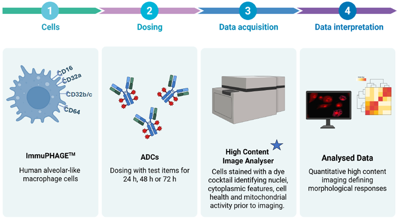
Image Credit: Image courtesy of Joel J et al., in partnership with ELRIG (UK) Ltd.
 Two assays recommended:
- Macrophage health and morphology assessment – morph_ONE assay5
- ADC uptake by macrophages assessment – ADC internalization assay
Results
Individual cell data analysis indicated morphological macrophage changes resulted from ADC or its component treatments
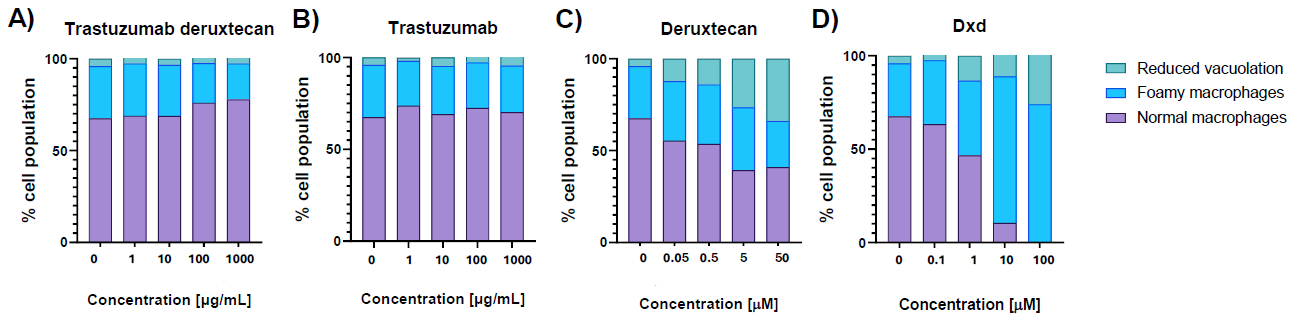
Figure 1. Individual cell analysis displaying phenotype distribution of ImmuPHAGE™ alveolar macrophages after 72 h exposure to trastuzumab-deruxtecan (A), trastuzumab (B), deruxtecan (C) and Dxd (D). Stacked bar charts represent the proportion of cells classified as normal macrophages (purple bars), foamy macrophages (blue bars), or reduced-vacuolation macrophages (green bars) at increasing concentrations. Briefly, cells were fixed and treated with a cocktail of fluorescent dyes staining for cellular features (nucleus, cytoplasm, and mitochondrial activity). Images were captured with a 40x objective in standard 2D imaging mode using the InCell Analyser 6000. Each sample was imaged using 16 fields, representing a total of 50 to 1000 cells per well. Images were analyzed using InCell Developer Toolbox 1.9.2 software and followed by generation of drug-induced phenotypes. Image Credit: Image courtesy of Joel J et al., in partnership with ELRIG (UK) Ltd.
Trastuzumab deruxtecan, trastuzumab, and the untreated group displayed similar phenotype profiles, with 67%-77% of cells showing normal morphology, 20-28% foamy macrophages, and 2%-5% of cells showing reduced vacuolation.
Treatment with deruxtecan (payload with linker) increased the number of cells with reduced vacuolation dose-dependently, from 4% untreated to 34% at 50 µM deruxtecan.
Dxd induced a dose-dependent increase in the number of foamy macrophages, from 20% untreated to 74% at>10 µM Dxd.
Detailed morphological analysis of foamy macrophages allowed identification and quantification of differences in foamy macrophage type
Foamy macrophage is an umbrella term for a macrophage that displays increased vacuolation.
Exposure to Dxd resulted in a 57-fold increase in the subpopulation of cells associated with low mitochondrial activity, smaller area, fewer vacuoles, and a smaller vacuolar area (Figure 2, blue bar).
The subpopulation associated with higher mitochondrial activity, larger cell area, and more vacuoles also increased 16-fold (Figure 2, dark purple bar).
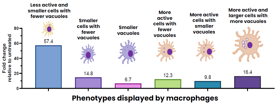
Figure 2. Fold change in phenotype distribution following 72 h exposure to 30 µM Dxd (payload). Each cell was assessed for 4 descriptors (mitochondrial activity, cell size, number of vacuoles, and size of vacuoles), categorized into levels (low, moderate, high) relative to untreated control cells, generating 81 potential phenotypes. Data is expressed as a fold change relative to the untreated cell baseline with fold changes > 5 depicted. Image Credit: Image courtesy of Joel J et al., in partnership with ELRIG (UK) Ltd.
Investigating ADC internalization
Phenotypic changes reflect macrophage responses but do not identify the route of payload uptake
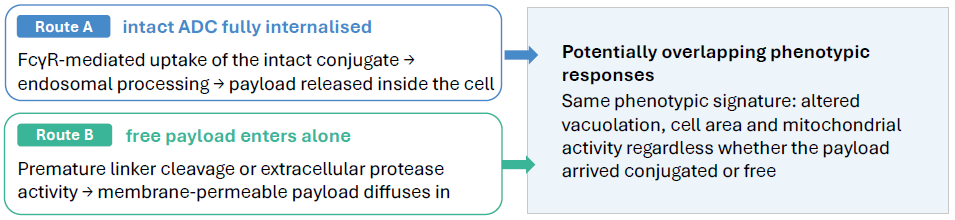
Image Credit: Image courtesy of Joel J et al., in partnership with ELRIG (UK) Ltd.
The internalization assay
Quantifying ADC uptake and intracellular localization in ImmuPHAGE™ could complement phenotypic profiling. FcγR blockade could help establish the contribution of receptor-mediated uptake to macrophage responses.
Implications for ADC development
These data could inform Fc or linker optimization and candidate selection to reduce off-target exposure. Prediction of clinical pulmonary toxicity would require further validation.
Conclusion and impact
High-content image analysis using morph_ONE™ provides a sensitive, human-relevant approach for detecting early macrophage responses to ADCs and their individual components.
Resolving phenotype at the single-cell level identifies altered vacuolation, cell area changes, and metabolic stress that population averages miss, and separates responses elicited by the intact conjugate from those driven by the payload alone.
Work is ongoing to determine whether uptake is FcγR-dependent, which connects this phenotype to an ADC design decision.
References
- Nguyen TD et al.(2023) Cancers (Basel); 15(3):713
- Colombo R et al. (2024) Cancer Discovery; 14(11): 2089 - 2108
- AbuhelwaZ et al. (2022) Drugs; 82(9):979–987
- Aoyama, M., et al. (2022). Fcγ receptor-dependent internalization and off-target cytotoxicity of antibody-drug conjugate aggregates. Pharmaceutical Research, 39(1), pp.89–103. DOI:10.1007/s11095-021-03158-x. https://link.springer.com/article/10.1007/s11095-021-03158-x.
- Hoffman, E., et al. (2023). Profiling alveolar macrophage responses to inhaled compounds using in vitro high content image analysis. Toxicology and Applied Pharmacology, 474, p.116608. DOI:10.1016/j.taap.2023.116608. https://linkinghub.elsevier.com/retrieve/pii/S0041008X23002478.
About ImmuONE Ltd.
ImmuONE specializes in human alveolar macrophage responses, epithelial cell culture platforms and testing services. Our respiratory tissue models closely resemble lung physiology to provide relevant, reliable, and reproducible results.
ImmuONE is partnered with the industry and academia to develop sustainable technologies for the future to support human-relevant, mechanistic driven safety assessments and develop animal product-free cell models. We offer a range of standard and bespoke assessments for inhalation safety assessment needs.
ImmuONE provides innovative solutions for inhalation toxicology, irritation sensitization, alveolar clearance, inflammation and can tailor to your inhalation safety assessment needs.
About ELRIG (UK) Ltd.
The European Laboratory Research & Innovation Group (ELRIG) is a leading European not-for-profit organization that exists to provide outstanding scientific content to the life science community. The foundation of the organization is based on the use and application of automation, robotics and instrumentation in life science laboratories, but over time, we have evolved to respond to the needs of biopharma by developing scientific programmes that focus on cutting-edge research areas that have the potential to revolutionize drug discovery.
Comprised of a global community of over 12,000 life science professionals, participating in our events, whether it be at one of our scientific conferences or one of our networking meetings, will enable any of our community to exchange information, within disciplines and across academic and biopharmaceutical organizations, on an open access basis, as all our events are free-of-charge to attend!
Our values
Our values are to always ensure the highest quality of content and that content will be made readily accessible to all, and that we will always be an inclusive organization, serving a diverse scientific network. In addition, ELRIG will always be a volunteer led organization, run by and for the life sciences community, on a not-for-profit basis.
Our purpose
ELRIG is a company whose purpose is to bring the life science and drug discovery communities together to learn, share, connect, innovate and collaborate, on an open access basis. We achieve this through the provision of world class conferences, networking events, webinars and digital content.
Sponsored Content Policy: News-Medical.net publishes articles and related content that may be derived from sources where we have existing commercial relationships, provided such content adds value to the core editorial ethos of News-Medical.net, which is to educate and inform site visitors interested in medical research, science, medical devices and treatments.
Last Updated: Oct 27, 2026