This article and associated images are based on a poster originally authored by Katarina Rydén Markinhuhta, Linda Hägerstrand, Margareta Behrendt, Weike Bao, Qing-Dong Wang, Alison Rowles and Pernilla Eliasson and presented at ELRIG Drug Discovery 2025 in affiliation with AstraZeneca.
This poster is being hosted on this website in its raw form, without modifications. It has not undergone peer review but has been reviewed to meet AZoNetwork's editorial quality standards. The information contained is for informational purposes only and should not be considered validated by independent peer assessment.

Introduction
Traditional assessment of cardiac hypertrophy in rodent models relies on stereological analysis at the cellular level, which provides high precision but is limited by complexity and low throughput.1
Macroscopic measures, such as heart weight to tibia length ratio (HW/TL) and left ventricular (LV) mass, are commonly used to measure cardiac hypertrophy but lack specificity, as increases in weight may reflect non-hypertrophic changes within the myocardium.
To overcome the limitations of conventional hypertrophic methods, the research team developed an AI-based image analysis algorithm for accurate and high-throughput quantification of cardiomyocyte hypertrophy using histological sections.
Study design and results
Experimental overview. Cardiomyocyte membranes in rodent heart tissue were visualized by laminin immunohistochemistry and scanned as virtual whole-slide images.
A deep learning algorithm, implemented in Visiopharm and trained on manually annotated reference images, was used to automatically identify cardiomyocytes with centrally located nuclei, ensuring selection of true cross-sectional profiles and minimizing sectioning artifacts. The average cardiomyocyte cross-sectional area (CM area) was used as a quantitative measure of hypertrophy.
Results. The method was validated in two established rodent models of hypertrophy: the Transverse Aortic Constriction (TAC) mouse model and the Zucker Diabetic Sprague-Dawley Fatty (ZSF1) rat model. In both, AI-based morphometric analysis detected hypertrophy with sensitivity equal to or greater than conventional metrics such as the HW/TL ratio and LV mass, which was cautiously reviewed by a pathologist.
Image analysis of CM area correlates significantly with HW/TL ratio and LV mass in hypertrophic disease models
Hypertrophy measurements in TAC mouse model
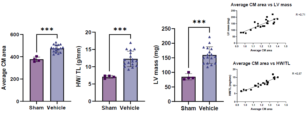
Image Credit: Image courtesy of Katarina Rydén Markinhuhta et al., in partnership with ELRIG (UK) Ltd.
Hypertrophy measurements in ZSF1 rat model
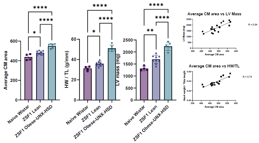
Image Credit: Image courtesy of Katarina Rydén Markinhuhta et al., in partnership with ELRIG (UK) Ltd.
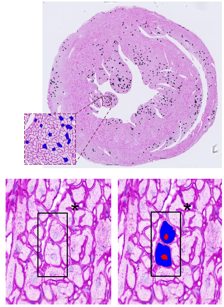
Cardiomyocytes, with cross-sectional profile, visualized with laminin immunohistochemistry and automated identification using algorithm. Image Credit: Image courtesy of Katarina Rydén Markinhuhta et al., in partnership with ELRIG (UK) Ltd.
Conclusions
The automated, AI-based image analysis of cardiomyocyte cross-sectional area presents a robust, specific, and scalable strategy for quantifying pathological cardiac hypertrophy in rodent models, surpassing conventional methods.
This platform enables detailed, high-throughput cell-level assessment of pathological cardiac hypertrophy, providing a useful tool for identifying new pharmacological treatments for cardiac hypertrophy.
References
- Mühlfeld, C., Nyengaard, J.R., and Mayhew, T.M. (2010). A review of state-of-the-art stereology for better quantitative 3D morphology in cardiac research. 19(2), pp.65–82. https://doi.org/10.1016/j.carpath.2008.10.015.
- Bensley, J.G., et al. (2016). Three-dimensional direct measurement of cardiomyocyte volume, nuclearity, and ploidy in thick histological sections. Scientific Reports, 6(1). https://doi.org/10.1038/srep23756.
About AstraZeneca
AstraZeneca is a global, science-led patient-focused pharmaceutical company that focuses on transforming the future of healthcare by unlocking the power of what science can do for people, society and the planet.
Science defines who we are. We push the boundaries of science to deliver life-changing medicines. It is why we come to work every day and is part of our DNA. We are committed to operating sustainably, in a way that recognises the interconnection between business growth, the needs of society and the limitations of our planet. We want the way we work to be inclusive, open and collaborative. This approach runs through all that we do.
About ELRIG (UK) Ltd.
The European Laboratory Research & Innovation Group (ELRIG) is a leading European not-for-profit organization that exists to provide outstanding scientific content to the life science community. The foundation of the organization is based on the use and application of automation, robotics and instrumentation in life science laboratories, but over time, we have evolved to respond to the needs of biopharma by developing scientific programmes that focus on cutting-edge research areas that have the potential to revolutionize drug discovery.
Comprised of a global community of over 12,000 life science professionals, participating in our events, whether it be at one of our scientific conferences or one of our networking meetings, will enable any of our community to exchange information, within disciplines and across academic and biopharmaceutical organizations, on an open access basis, as all our events are free-of-charge to attend!
Our values
Our values are to always ensure the highest quality of content and that content will be made readily accessible to all, and that we will always be an inclusive organization, serving a diverse scientific network. In addition, ELRIG will always be a volunteer led organization, run by and for the life sciences community, on a not-for-profit basis.
Our purpose
ELRIG is a company whose purpose is to bring the life science and drug discovery communities together to learn, share, connect, innovate and collaborate, on an open access basis. We achieve this through the provision of world class conferences, networking events, webinars and digital content.
Sponsored Content Policy: News-Medical.net publishes articles and related content that may be derived from sources where we have existing commercial relationships, provided such content adds value to the core editorial ethos of News-Medical.Net which is to educate and inform site visitors interested in medical research, science, medical devices and treatments.
Last Updated: Nov 13, 2025
 Aptamer Group and AstraZeneca to develop new drug delivery vehicles