This article and associated images are based on a poster originally authored by Mayur S. Kale, Benjamin J. Small, Ogechi Nkeonye, Shalini Kumarasamy, Mohit Joshi, Devshree Parganiha, Kate Butcher, Christopher Heeschen, Yaohe Wang, Mohammad Najlah, Vinodh Kannappan and Weiguang Wang and presented at ELRIG Drug Discovery 2026 in affiliation with University of Wolverhampton, Disulfican Ltd, Vellore Institute of Technology, Shanghai Jiao Tong University, Queen Mary University of London, Anglia Ruskin University and University of Liverpool.
This poster is being hosted on this website in its raw form, without modifications. It has not undergone peer review but has been reviewed to meet AZoNetwork's editorial quality standards. The information contained is for informational purposes only and should not be considered validated by independent peer assessment.

Background and rationale
Pancreatic ductal adenocarcinoma (PDAC) remains difficult to treat because of late diagnosis, limited therapeutic response, and acquired drug resistance. Drug repurposing provides a cost- and time-efficient strategy to identify new anticancer therapies from clinically established medicines with known safety profiles.
Disulfiram (DSF) is an anti-alcoholism drug with emerging anticancer potential in vivo. DSF is rapidly metabolized to diethyldithiocarbamate (DDC), which chelates transition metals such as copper or zinc to form cytotoxic metal-DDC complexes.
Albumin-formulated Alb-CuDDC and Alb-ZnDDC were developed to improve the solubility, stability, and cellular delivery of these complexes. Both formulations are cytotoxic in PANC-1 PDAC cells; however, they appear to act through distinct mechanisms. Previous work showed that Alb-CuDDC induces nuclear aggregation of NPL4, leading to proteotoxic stress, whereas the mechanism underlying Alb-ZnDDC-induced cytotoxicity remains unclear.
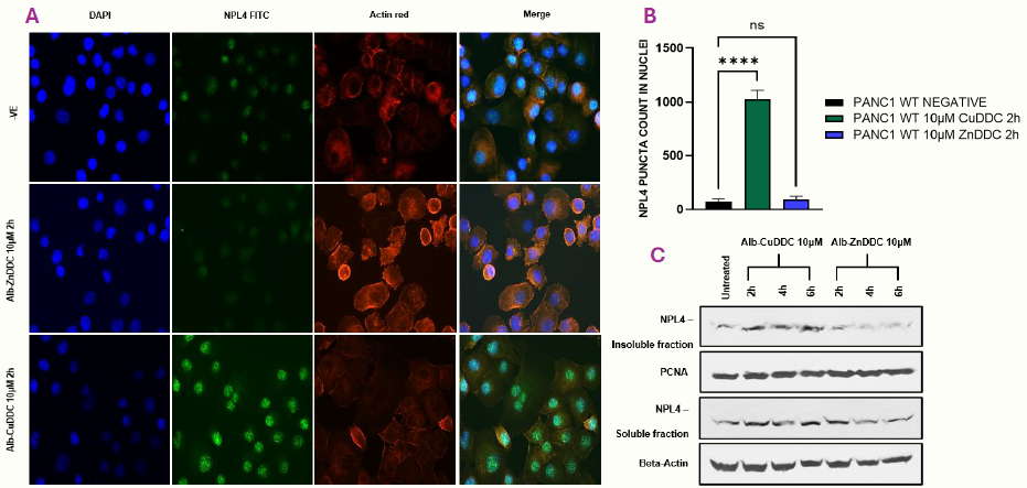
Figure 1. Alb-CuDDC, but not Alb-ZnDDC, induces nuclear NPL4 aggregation in parental PANC-1 cells. (A) Representative immunofluorescence images showing DAPI (blue), NPL4 (green) and actin (red). (B) Quantification of nuclear NPL4 puncta count showing significant NPL4 aggregation after treatment with Alb-CuDDC, but not with Alb-ZnDDC. (C) Western blot analysis of NPL4 expression in PANC-1 cells treated with Alb-CuDDC and Alb-ZnDDC. Image Credit: Image courtesy of Mayur S. Kale et al., in partnership with ELRIG (UK) Ltd.
To compare the anticancer mechanisms of Alb-CuDDC and Alb-ZnDDC in PANC-1 cells and investigate mechanisms underlying acquired resistance to Alb-ZnDDC:
- Compare the mechanisms of Alb-CuDDC and Alb-ZnDDC in PANC-1 cells
- Investigate whether Alb-CuDDC and Alb-ZnDDC induce lysosomal membrane permeabilization (LMP)
- Characterize ZR6 resistance adaptations in PANC-1 ZR6 cells
- Determine whether CST2 contributes to lysosomal protection and Alb-ZnDDC resistance
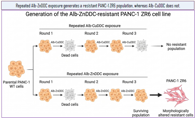
Figure 2. Generation of Alb-ZnDDC-resistant PANC-1 cells by repeated drug exposure. Created with BioRender.com. Image Credit: Mayur S. Kale et al., in partnership with ELRIG (UK) Ltd.
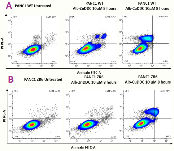
Figure 3. Representative Annexin V/PI flow cytometry plots of apoptosis in (A) parental PANC-1 WT and (B) AlbZnDDC-resistant PANC-1 ZR6 cells following treatment with AlbZnDDC or Alb-CuDDC (10 µM, 8 h). Quadrants indicate viable, early apoptotic, late apoptotic, and necrotic cells. Image Credit: Mayur S. Kale et al., in partnership with ELRIG (UK) Ltd.
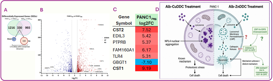
Figure 4. RNA sequencing identifies candidate genes associated with Alb-ZnDDC resistance and proposes a mechanistic hypothesis. (A) Venn diagram of differentially expressed genes (DEGs) in Alb-ZnDDC-resistant PANC-1 ZR6 cells. (B) Volcano plot showing significantly dysregulated genes. (C) Top differentially expressed genes, including marked upregulation of CST1 and CST2. (D) Proposed mechanism whereby Alb-ZnDDC induces lysosomal membrane permeabilization (LMP), cathepsin release and apoptosis, while CST1/CST2 overexpression in ZR6 cells may inhibit cathepsin activity and contribute to resistance. Image Credit: Mayur S. Kale et al., in partnership with ELRIG (UK) Ltd.
Alb-ZnDDC, but not Alb-CuDDC, induces LMP in parental PANC -1 WT cells
- Alb-ZnDDC reduced LysoTracker-positive puncta (Fig. 5A) and increased galectin-3 puncta/galectin-3-LAMP1 co-localization (Fig. 5B & E), consistent with LMP
- LAMP1/2 protein changes (Fig. 5D) and increased cytosolic cathepsin activity (Fig. 5F) further supported lysosomal stress and leakage after Alb-ZnDDC treatment
- FluoZin imaging showed increased zinc-associated intracellular signal following Alb-ZnDDC exposure (Fig. 5C)
- In contrast, Alb-CuDDC caused minimal lysosomal disruption and no comparable LMP markers across Fig. 5A–F, supporting distinct intracellular mechanisms.
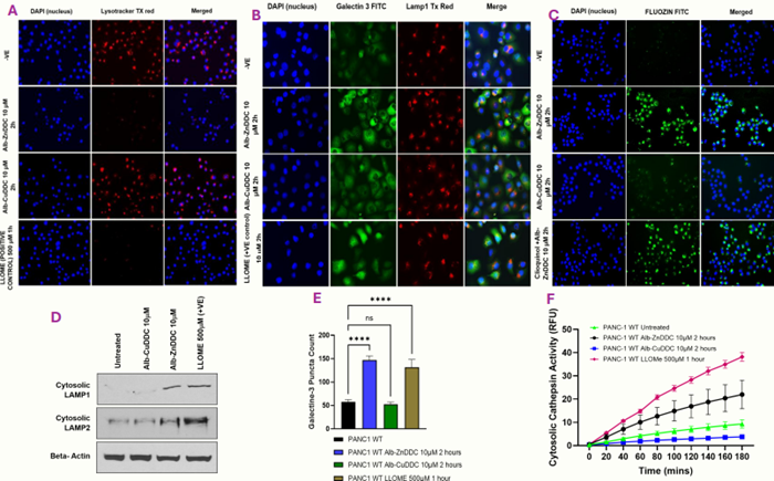
Figure 5. Alb-ZnDDC induces lysosomal membrane permeabilization (LMP) in parental PANC-1 cells. (A) LysoTracker loss indicates lysosomal disruption. (B) Galectin-3 puncta and galectin-3/LAMP1 co-localization indicate lysosomal damage. (C) FluoZin imaging shows increased zinc-associated intracellular signal following Alb-ZnDDC treatment. (D) Cytosolic LAMP1/2 protein changes support lysosomal stress. (E) Galectin-3 puncta were quantified. (F) Increased cytosolic cathepsin activity indicates lysosomal leakage and cathepsin release. Data in (E) are presented as mean ± SEM from n = 4 independent experiments. Statistical analysis was performed using one-way ANOVA with Tukey’s multiple-comparisons test; ns, not significant; ***p < 0.0001. Image Credit: Mayur S. Kale et al., in partnership with ELRIG (UK) Ltd.
Alb-ZnDDC resistance in PANC-1 ZR6 cells
- As shown in Fig. 6A, C, F and G, ZR6 cells preserved LysoTracker-positive puncta and displayed reduced cytosolic cathepsin activity following Alb-ZnDDC exposure, consistent with attenuated LMP
- FluoZin-positive signal remained increased in ZR6 cells following Alb-ZnDDC exposure (Fig. 6B and H), indicating that ZR6 cells can accommodate elevated intracellular zinc-associated signal
- Together, these adaptations contribute to Alb-ZnDDC resistance in ZR6 cells
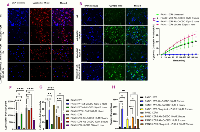
Figure 6. PANC-1 ZR6 cells exhibit reduced Alb-ZnDDC-induced lysosomal damage and altered stress-adaptation markers. (A–B) Annexin V/PI analysis shows reduced cell death in ZR6 cells following Alb-ZnDDC treatment. (C, F) LysoTracker imaging and quantification indicate preservation of lysosomal puncta in ZR6 cells. (D, G) Cytosolic cathepsin activity is reduced in ZR6 cells following Alb-ZnDDC exposure. (E, H) FluoZin imaging and quantification show retained zinc-associated intracellular signal in ZR6 cells. (I–N) RT-qPCR analysis shows altered metallothionein gene expression in ZR6 cells. Data are presented as mean ± SEM. Statistical significance is shown on the graphs: ns, *p < 0.05; **p < 0.01; ***p < 0.001; ****p < 0.0001. Statistical comparisons were performed using one-way ANOVA with Tukey’s multiple-comparisons test. Image Credit: Mayur S. Kale et al., in partnership with ELRIG (UK) Ltd.
CST2 partially increases Alb-ZnDDC tolerance
- CST2 was elevated in ZR6 cells and successfully overexpressed in C10 and C39 PANC-1 clones, as confirmed by RT-PCR and western blotting (Fig. 7A, B and D)
- Whole-cell cathepsin activity was reduced in CST2-overexpressing clones compared with mock cells (Fig. 7C )
- Cytosolic cathepsin activity was attenuated in CST2-overexpressing clones following Alb-ZnDDC or LLOMe exposure (Fig. 8A–C)
- Tolerance to Alb-ZnDDC and LLOMe increased in CST2-overexpressing clones, with C10 showing the greatest Alb-ZnDDC resistance. Alb-CuDDC sensitivity was unchanged (Fig. 8D–F)
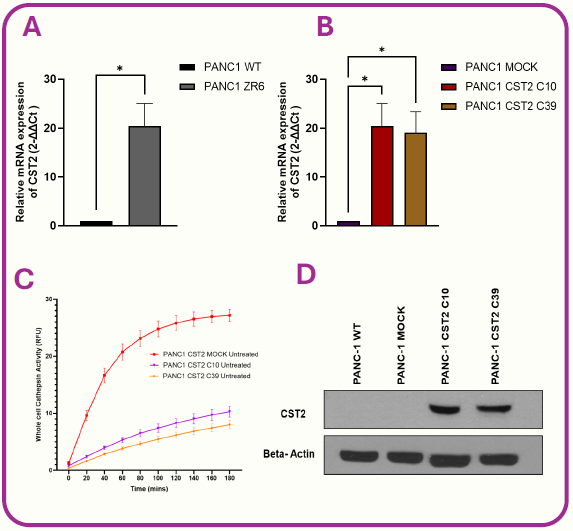
Figure 7. Validation of CST2-overexpressing PANC-1 clones. (A–B) CST2 mRNA expression in ZR6 cells and CST2 C10/C39 clones. (C) Whole-cell cathepsin activity in mock and CST2- overexpressing clones. (D) Western blot confirmation of CST2 protein expression; β-actin, loading control. Data are mean ± SEM, n = 4; *p < 0.05. Image Credit: Mayur S. Kale et al., in partnership with ELRIG (UK) Ltd.
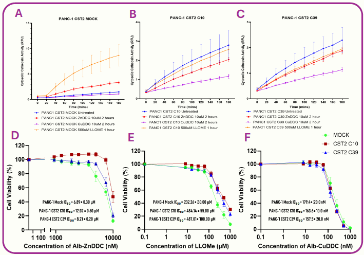
Figure 8. CST2 overexpression partially protects PANC-1 cells from Alb-ZnDDC- and lysosomal stress. (A–C) Cytosolic cathepsin activity in mock, CST2 C10 and CST2 C39 cells following AlbZnDDC, Alb-CuDDC or LLOMe treatment. (D–F) Cell-viability curves following exposure to AlbZnDDC, LLOMe or Alb-CuDDC. Image Credit: Mayur S. Kale et al., in partnership with ELRIG (UK) Ltd.
Discussion, conclusion and future work

Figure 9. RT-qPCR analysis of metallothionein and metal-transporter gene expression in PANC-1 WT and Alb-ZnDDC-resistant ZR6 cells. Relative mRNA expression of (I) MT1M, (J) MT1X, (K) MT1F, (L) MT1E, (M) SLC30A1 and (N) SLC31A1 was assessed in untreated and Alb-ZnDDC-treated (7 µM) PANC-1 WT and ZR6 cells. Gene expression was calculated using the 2^−ΔΔCt method. Data are presented as mean ± SEM. Statistical analysis was performed using one-way ANOVA with Tukey’s multiple-comparisons test; ns, not significant; **p < 0.01; ***p < 0.001. Image Credit: Mayur S. Kale et al., in partnership with ELRIG (UK) Ltd.
Conclusion
- Alb-CuDDC and Alb-ZnDDC are cytotoxic in PANC-1 cells but act through distinct intracellular mechanisms: Alb-CuDDC induces nuclear NPL4 aggregation consistent with CuET-associated proteotoxic stress, whereas Alb-ZnDDC induces zinc-associated LMP, cathepsin release, and apoptosis
- Acquired Alb-ZnDDC resistance in PANC-1 ZR6 cells is associated with lysosomal protection, altered zinc handling, reduced cathepsin release, and increased CST2 expression
- CST2 overexpression reduced cathepsin activity and partially increased tolerance to Alb-ZnDDC and LLOMe, but not Alb-CuDDC
Defining and targeting Alb-ZnDDC resistance mechanisms
- Validate CST2 and identify additional ZR6 resistance pathways, including zinc-buffering, ROS adaptation, and lysosomal repair
- Evaluate Alb-ZnDDC and Alb-CuDDC in vivo for efficacy, toxicity, and resistance development
References
- Skrott, Z., et al. (2017). Alcohol-abuse drug disulfiram targets cancer via p97 segregase adaptor NPL4. Nature, 552(7684), pp.194–199. DOI:10.1038/nature25016. https://www.nature.com/articles/nature25016
- Small, B.J. (2025) In vitro investigation of the anticancer activity and molecular mechanisms of action of albumin nano-encapsulated Cu(DDC)2 and Zn(DDC)2 in human pancreatic cancer cells. PhD thesis. University of Wolverhampton.
- Boya, P. and Kroemer, G. (2008). Lysosomal membrane permeabilization in cell death. Oncogene, 27(50), pp.6434–6451. DOI:10.1038/onc.2008.310. https://www.nature.com/articles/onc2008310
- Jia, J., et al. (2020). Galectin-3 coordinates a cellular system for lysosomal repair and removal. Developmental Cell, 52(1), pp.69–87.e8. DOI:10.1016/j.devcel.2019.10.025. https://www.cell.com/developmental-cell/fulltext/S1534-5807(19)30903-7
Acknowledgments
We thank staff and colleagues at the RIHS for their technical support. We thank the University of Wolverhampton and Disulfiram Ltd for providing research facilities and support. We thank the ELRIG for providing travel grant support to present this work at this conference.
 About University of Wolverhampton
Opportunities for students have been at the heart of our mission for over 190 years.
The University of Wolverhampton’s roots go back to the 19th century Wolverhampton Mechanics’ Institutes and School of Art. It offers a broad range of subjects at foundation, undergraduate and postgraduate level – many accredited by prestigious professional bodies. Over 21,000 students studying at three main teaching campuses in the Midlands and partners nationwide benefit from innovative and excellent teaching from a QAA-commended university listed in the top ten universities for social mobility in a paper published by the Higher Education Policy Institute, with world-class research and a 87% graduate employability rate (Graduate Outcomes report 2021).
About ELRIG (UK) Ltd.
The European Laboratory Research & Innovation Group (ELRIG) is a leading European not-for-profit organization that exists to provide outstanding scientific content to the life science community. The foundation of the organization is based on the use and application of automation, robotics, and instrumentation in life science laboratories, but over time, we have evolved to respond to the needs of biopharma by developing scientific programs that focus on cutting-edge research areas that have the potential to revolutionize drug discovery.
Comprised of a global community of over 12,000 life science professionals, participating in our events, whether it be at one of our scientific conferences or one of our networking meetings, will enable any of our community to exchange information, within disciplines and across academic and biopharmaceutical organizations, on an open access basis, as all our events are free of charge to attend!
Our values
Our values are to always ensure the highest quality of content, that content will be made readily accessible to all, and that we will always be an inclusive organization, serving a diverse scientific network. In addition, ELRIG will always be a volunteer-led organization, run by and for the life sciences community, on a not-for-profit basis.
Our purpose
ELRIG is a company whose purpose is to bring the life science and drug discovery communities together to learn, share, connect, innovate, and collaborate on an open-access basis. We achieve this through the provision of world-class conferences, networking events, webinars, and digital content.
Sponsored Content Policy: News-Medical.net publishes articles and related content that may be derived from sources where we have existing commercial relationships, provided such content adds value to the core editorial ethos of News-Medical.net, which is to educate and inform site visitors interested in medical research, science, medical devices, and treatments.
Last Updated: Oct 29, 2026