This article and associated images are based on a poster originally authored by Aida Ashrafzadeh, Marcos Rubio-Alarcón, Graham Ladds, and Taufiq Rahman, and presented at ELRIG Drug Discovery 2025 in affiliation with the University of Cambridge.
This poster is being hosted on this website in its raw form, without modifications. It has not undergone peer review but has been reviewed to meet AZoNetwork's editorial quality standards. The information contained is for informational purposes only and should not be considered validated by independent peer assessment.

Introduction
Store-operated calcium entry (SOCE) is triggered when the endoplasmic reticulum (ER) calcium stores are depleted, activating calcium release–activated calcium (CRAC) channels.1
This calcium influx not only refills ER stores but also drives activation of the NFAT signalling pathway, a key regulator of immune and neuronal gene expression programs.
CRAC channels are well-established therapeutic targets. Several small-molecule inhibitors have been developed for peripheral inflammatory and autoimmune diseases.
Increasing evidence links CRAC channel upregulation to CNS disorders, including autoimmune encephalomyelitis, neurodegeneration, neuropathic pain, and brain cancers.2 However, brain-permeable CRAC inhibitors remain scarce.
This study aims to identify and characterize brain-penetrant SOCE inhibitors using an integrated computational and experimental approach.
Ligand-based virtual screening
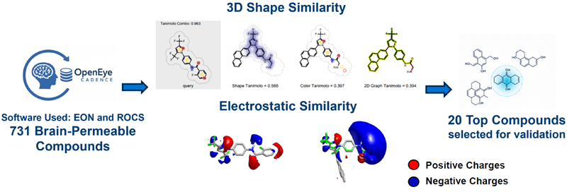
Image Credit: Image courtesy of Aida Ashrafzadeh et al., in partnership with ELRIG (UK) Ltd.
Ca2+ influx assay
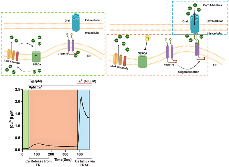
Figure 1. Stepwise illustration of SOCE activation during the calcium flux assay using FlexStation system in jurkat T cells. Image Credit: Aida Ashrafzadeh et al., in partnership with ELRIG (UK) Ltd.
- Basal state: ER Ca2+ levels are maintained by SERCA pumps, balancing passive Ca2+ leak.
- Store depletion: SERCA inhibition by thapsigargin (Tg) leads to ER Ca2+ depletion, activating STIM1/2, which gate Orai1 channels.
- SOCE activation: Upon introducing extracellular Ca2+, the ions flow into the cells through Orai channels
Ca2+ influx screening
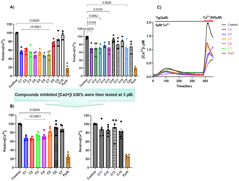
Figure 2. A) Bar diagrams represent the relative peak Ca2+ entry [Ca2+]i. Compounds initially tested at 10 μM. B) Compounds with ≥20 % inhibition were further explored for dose response assays. C) Representative Tg-evoked Ca2+ signal traces for C1-C4. Image Credit: Image courtesy of Aida Ashrafzadeh et al., in partnership with ELRIG (UK) Ltd.
Pairwise comparisons are shown relative to the control. Pyr6 vs. control: p < 0.001 (applies to all figures).
C1-C4 dose-response curve
![A)</strong> Effect of C1–C4 Compounds at different concentrations on SOCE.<strong> B)</strong> Concentration–response curves of C1-C4 showing their inhibitory effect on SOCE. <strong>C)</strong> Graph shows effect of C1-C4 on Ca<sup>2+</sup> release from ER ([Ca<sup>2+</sup>]<sub>ER</sub>). C1–C3 showed no significant effects at any concentration, while C4 inhibited [Ca<sup>2+</sup>]<sub>ER</sub> at 3, 10 and 30 μM](https://www.news-medical.net/images/Article_Images/ImageForArticle_26772_17623425143337045.png)
Figure 3. A) Effect of C1–C4 Compounds at different concentrations on SOCE. B) Concentration–response curves of C1-C4 showing their inhibitory effect on SOCE. C) Graph shows effect of C1-C4 on Ca2+ release from ER ([Ca2+]ER). C1–C3 showed no significant effects at any concentration, while C4 inhibited [Ca2+]ER at 3, 10 and 30 μM. Image Credit: Image courtesy of Aida Ashrafzadeh et al., in partnership with ELRIG (UK) Ltd.
NFAT reporter assay
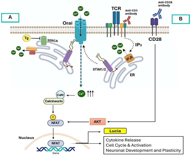
Figure 4. Schematic representation of NFAT pathway activation and reporter assay principle. TCR/CD28 activation (A) or Tg-induced ER Ca2+ depletion (B) triggers SOCE, activating calcineurin–NFAT signalling. NFAT translocates to the nucleus and induces Lucia reporter expression, measured as luminescence. Image Credit: Image courtesy of Aida Ashrafzadeh et al., in partnership with ELRIG (UK) Ltd.
C1-C3 NFAT assay
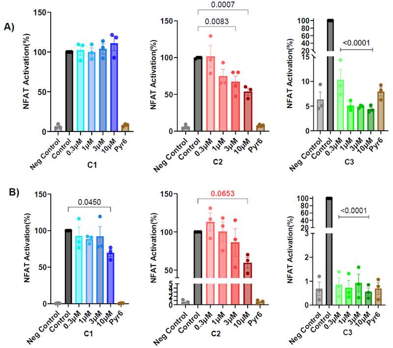
Figure 5. NFAT reporter assay following stimulation by TCR activation (A) or Tg (B) in Jurkat-Lucia™ NFAT-CD28 reporter cells treated with C1-C3. Image Credit: Image courtesy of Aida Ashrafzadeh et al., in partnership with ELRIG (UK) Ltd.
NFAT activation was quantified as a percentage relative to the stimulated control group. Pyr6 and negative control vs. control: p < 0.001 (applies to all figures).
Conclusion
- Functional assays confirmed the robustness of the ligand-based virtual screening pipeline, yielding one compound that inhibits CRAC with an IC50 in the nanomolar range (C2) and three compounds with IC50s in the micromolar range (C1, C3, C4).
- C3 strongly inhibited NFAT under both TCR- and Tg-mediated activation despite moderate SOCE inhibition, while C1 and C2 did not suppress NFAT at concentrations where they inhibited CRAC. It suggests that CRAC inhibition may not always suppress NFAT, possibly due to compensatory Ca2+ signals maintaining intracellular Ca2+ levels or actions on other regulators.
- C4 showed toxicity in the six-hour NFAT assay and inhibited Ca2+ release from ER, so it was not further characterized.
- C3, with its strong NFAT suppression, may be advantageous in neuroinflammatory diseases, while C1 and C2, with weaker effects, might be better suited for conditions where immune function should be preserved.
Future work will focus on further characterizing the most promising hits, investigating their NFAT-related mechanisms in greater detail, and testing them in brain cancer models to establish their translational relevance.
References
- Prakriya, M. and Lewis, R.S. (2015). Store-Operated Calcium Channels. Physiological Reviews, 95(4), pp.1383–1436. DOI: 10.1152/physrev.00020.2014. https://journals.physiology.org/doi/full/10.1152/physrev.00020.2014.
- Feske, S. (2019). CRAC channels and disease – From human CRAC channelopathies and animal models to novel drugs. Cell Calcium, 80, pp.112–116. DOI: 10.1016/j.ceca.2019.03.004. https://www.sciencedirect.com/science/article/abs/pii/S0143416019300478?via%3Dihub
About the University of Cambridge 
The University of Cambridge is one of the world's foremost research universities.
The University is made up of 31 Colleges and over 150 departments, faculties, schools and other institutions. Its mission is 'to contribute to society through the pursuit of education, learning, and research at the highest international levels of excellence'.
About ELRIG (UK) Ltd.
The European Laboratory Research & Innovation Group (ELRIG) is a leading European not-for-profit organization that exists to provide outstanding scientific content to the life science community. The foundation of the organization is based on the use and application of automation, robotics and instrumentation in life science laboratories, but over time, we have evolved to respond to the needs of biopharma by developing scientific programs that focus on cutting-edge research areas that have the potential to revolutionize drug discovery.
Comprised of a global community of over 12,000 life science professionals, participating in our events, whether it be at one of our scientific conferences or one of our networking meetings, will enable any of our community to exchange information, within disciplines and across academic and biopharmaceutical organizations, on an open access basis, as all our events are free-of-charge to attend!
Our values
Our values are to always ensure the highest quality of content and that content will be made readily accessible to all, and that we will always be an inclusive organization, serving a diverse scientific network. In addition, ELRIG will always be a volunteer led organization, run by and for the life sciences community, on a not-for-profit basis.
Our purpose
ELRIG is a company whose purpose is to bring the life science and drug discovery communities together to learn, share, connect, innovate and collaborate, on an open access basis. We achieve this through the provision of world class conferences, networking events, webinars and digital content.
Sponsored Content Policy: News-Medical.net publishes articles and related content that may be derived from sources where we have existing commercial relationships, provided such content adds value to the core editorial ethos of News-Medical.Net which is to educate and inform site visitors interested in medical research, science, medical devices and treatments.
Last Updated: Nov 25, 2025