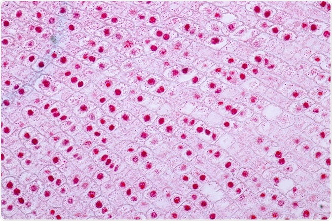
The adhesion of a cell to a surface is essential for many cellular processes, including the cell cycle. This article will describe recent studies which have expanded our knowledge of the role of focal adhesions in the cell cycle.
Skip to:
 Rattiya Thongdumhyu | Shutterstock
Recent research
Two recent studies (Dix et al., 2018 and Lock et al., 2018) have described how mitotic cells adhere to the extracellular matrix throughout the cell cycle.
Dix and colleagues found that the loss of the proteins paxillin, talin, and talin zyxin were linked to cellular rounding, and focal adhesion disassembly. Interestingly, the integrins and basal adhesion complexes at these sites remained active during the cell cycle, implicating them as key mediators of mitosis.
Lock et al. further identified a novel adhesion complex that was net-like in structure and is now known as reticular adhesions. Reticular adhesions are predominantly mediated by a specific type of integrin, αVβ5. These integrins are unique in that they are able to function independently. αVβ5 integrins may, however, interact with a distinct group of proteins that possess their own morphology: talin, paxillin, and zyxin.
How do focal adhesions change during the cell cycle?
The work of Jones et al. (2018) has provided insight into how progression through the cell cycle affects focal adhesion assembly and disassembly. They observed through an increase in the in 2D area of the focal adhesion when the cell progressed from the G1 to the S phase. This coincides with cell growth.
A peak area was subsequently recorded at the end of the S phase, overlapping with the beginning of G2, after which the paxillin stained area decreased. In addition to fluctuating area changes, the authors observed changes in focal adhesion location–paxillin stained focal adhesions were located at the cell periphery on transitioning from the G1 to S phase.
Distribution expanded to include the central and peripheral during the S phase, in agreement with the maximal area of the focal adhesion during the latter end of S phase/early G2. Degradation of focal adhesion during the transition from S to G2 phase was confirmed by live-cell microscopy, and by the end of G2, only peripheral focal adhesions remained.
Cell-cycle-dependent regulation of focal adhesions
To investigate the cell-cycle dependency of focal adhesion distribution and magnitude, Jones et al. investigated the role of cyclin‐dependent kinases (CDKs) in the regulation of adhesion dynamics. Earlier studies have implicated their role in phase transitions of the cell cycle.
Jones et al. inhibited CDK1 activity in asynchronous cells (cells at different phases of the cell cycle) and found that this reduced the area of focal adhesions. Subsequent work by Lock et al. confirmed the role of CDK1 as the driver of focal adhesion assembly, expansion, and distribution during S phase. This finding is counter-intuitive as CDK1 drives entry into the M phase, where cells assume a rounded morphology dissonant with up increased spread.
CDKs
CDKs function by pairing with regulatory subunits, called cyclins, in a specific manner; CDK1 pairs with the A and B type cyclins. Cyclin A2 is a subtype present in mammalian cells where it is increasingly expressed during the S phase and degraded on prometaphase entry. The subtype cyclin B1 displays a different expression pattern, appearing at the end of the S phase and persisting through prometaphase, through to late metaphase.
CDK1 targets
CDK1 targets are responsible for S-phase focal adhesion assembly include several actin‐associating proteins, particularly the formin-like 2 protein (FMNL2). FMNL2 is notable as it has been previously implicated in actin filament assembly. CDK1 phosphorylation of FMNL2 increases the adhesion area, its dephosphorylation in the G2 phase was implicated in decreased focal adhesion area and number. This was supported by an observed decrease in the central focal adhesions in cells that overexpressed FMNL2 mutant which was phosphorylation-adverse.
Jones et al. have provided some insight into targets of cell cycle kinases but the role FMNL2 regulates in cell cycle‐regulated focal adhesion formation and disassembly remains to be fully elucidated. Together with the work of Lock et al., there is evidence of additional effectors downstream of CDK1 that are involved in promoting adhesion in the S phase.
Sources
- Dix C. L., et al. (2018). The Role of Mitotic Cell-Substrate Adhesion Re-modeling in Animal Cell Division. Development Cell. doi.org/10.1016/j.devcel.2018.03.009.
- Jones, M. C., et al. (2018). Cell adhesion is regulated by CDK1 during the cell cycle. Journal of Cell Biology. 10.1083/jcb.201802088.
- Lock, J. G., et al. (2018). Reticular adhesions are a distinct class of cell-matrix adhesions that mediate attachment during mitosis. Nature Cell Biology. www.nature.com/articles/s41556-018-0220-2.
- Assoian, R. K. & Schwartz, M. A. (2001). Coordinate signaling by integrins and receptor tyrosine kinases in the regulation of G1 phase cell-cycle progression. Current Opinion in Genetics & Development. doi.org/10.1016/S0959-437X(00)00155-6
- Kiema, T., et al. (2006). The Molecular Basis of Filamin Binding to Integrins and Competition with Talin. Molecular Cell. doi.org/10.1016/j.molcel.2006.01.011.
Further Reading
Last Updated: Apr 4, 2023