Introduction
Scientific Background and Mechanisms
Evidence-Based Benefits of Ecosystem-Level Interventions
Why Spatial Context and Technologies Matter
Limitations and Conclusion
References
Further reading
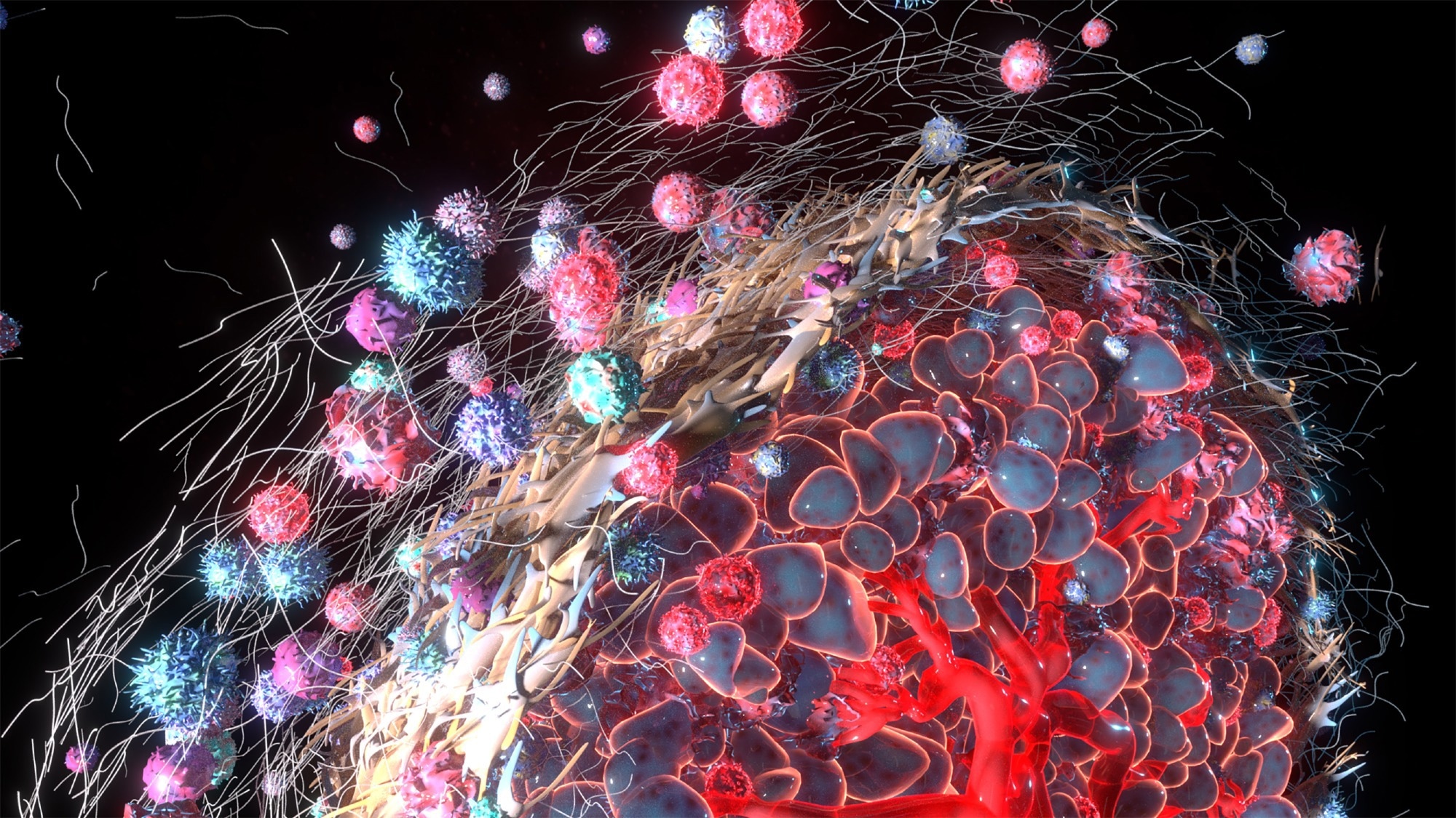
Cancer behaves as a complex, evolving system in which interactions between malignant cells and their surrounding environment shape growth, adaptation, and resistance to therapy. This understanding emphasizes the need to target the structural, spatial, and signaling networks that sustain the disease, enabling strategies that reshape its supportive environment rather than focusing solely on cell elimination.
 Study: Alpha Tauri 3D Graphics/Shutterstock.com
Since its origins in the late 18th century and formalization in 1907, oncology has largely focused on the discovery of the "magic bullet", a therapeutic agent capable of selectively eliminating malignant cells while sparing the host. Subsequent research, however, established that cancer cell lines are extraordinarily resilient to therapeutic interventions and often demonstrate disease recurrence after treatment ends, necessitating a fundamental re-evaluation of cancer’s nature.
Emerging research now indicates that a malignant tumor is not merely a collection of autonomous rogue cells but a complex, self-sustaining ecosystem, popularly termed the “onco-sphere”, a concept that describes multi-scale tumor-host interactions extending beyond the local microenvironment into systemic niches.2 This ecosystem has been observed to function as a dynamic system in which malignant cells interact with a diverse array of non-malignant host cells, extracellular matrices, and systemic physiological processes to ensure survival and expansion. This includes biomechanical, metabolic, and spatially organized interactions that collectively regulate tumor behavior.8
Take This Research With You. Download the Free PDF Version for Offline Reading By Clicking Here
Introduction
For decades, the dominant model in oncology focused on cell-autonomous features, primarily the genetic mutations that drive uncontrolled proliferation. However, the relatively recent scientific realization that cancer cells function as "living organisms" within a host habitat has led to the development of the cancer ecosystem framework, in which tumors are increasingly viewed as pseudo-organs composed of both malignant and non-malignant cell populations. Architecture and tissue organization play critical roles in determining malignancy, drug resistance, and disease progression.1,5,8
This conceptualization of cancer as an ecological and evolutionary process is considered one of the most significant shifts in modern medicine. Within this model (the cancer ecosystem framework), the tumor is viewed as a heterogeneous population undergoing clonal evolution, driven by both genetic diversity and environmental selective pressures within the tumor niche, as well as spatial constraints and resource gradients that shape evolutionary trajectories.1,5,8
Scientific Background and Mechanisms
The biochemistry of the cancer ecosystem is defined by intricate and mechanistically complex crosstalk between malignant cells and their surrounding matrix ("soil"). Recent research indicates that this crosstalk is governed by niche construction, where cancer cells modify their surrounding microenvironment to favor their survival, including metabolic reprogramming and immune modulation, as well as mechanical remodeling of extracellular matrices that alter tissue stiffness and signaling pathways.2,3,8
The Tumor Microenvironment
Video credit: BristolMyersSquibb/Youtube.com
High-resolution investigations have shown that the tumor microenvironment (TME) is composed primarily of cellular elements, including tumor-associated macrophages (TAMs), endothelial cells, pericytes, and diverse immune populations. Cellular elements, such as Cancer-Associated Fibroblasts (CAFs), have been shown to synthesize the extracellular matrix (ECM) and secrete growth factors, including Hepatocyte Growth Factor (HGF) and Fibroblast Growth Factor (FGF), thereby promoting tumor proliferation, invasion, and therapeutic resistance. Notably, CAFs frequently overexpress Fibroblast Activation Protein (FAP), a membrane-bound protease present in up to 90% of epithelial tumors, which contributes to ECM remodeling, angiogenesis, and immunosuppression.3,4,11
TAMs simultaneously promote angiogenesis via Vascular Endothelial Growth Factor (VEGF) signaling. Together, these processes create a microenvironment conducive to tumor growth and survival while also contributing to immune suppression through cytokine secretion and immune cell reprogramming. Anti-angiogenic therapies targeting VEGF pathways further demonstrate that disrupting these ecosystem-level interactions can significantly impair tumor progression.3,4,7
Emergent biochemical research indicates that the behavior of this ecosystem is driven by complex signaling loops, particularly involving Transforming Growth Factor (TGF-β), Interleukins (IL-6), and various chemokines. Studies have found that TGF-β serves as a critical mediator of the epithelial-to-mesenchymal transition (EMT), thereby allowing tumor cells to promote metastasis and resistance to apoptosis, while VEGF and hypoxia-inducible pathways further enhance angiogenesis and metastatic dissemination3,4.
Furthermore, CAFs can secrete CXCL12, which physically excludes host T-cells from the tumor core, thereby creating an "immune-excluded" phenotype and contributing to resistance to immune checkpoint blockade therapies. In this context, cancer stem cells (CSCs) actively remodel the TME to create protective, hypoxic niches that shield them from conventional cytotoxic therapies while simultaneously driving immune evasion and tumor relapse. These CSC–TME interactions are increasingly recognized as central drivers of therapy resistance and disease recurrence3-5.
Evidence-Based Benefits of Ecosystem-Level Interventions
The recent and ongoing transition of oncological practice from targeting individual cells to modulating the broader ecosystem has already demonstrated substantial benefits in preclinical and pilot clinical trials. Kang and colleagues (2026) highlight Anaplastic Thyroid Cancer (ATC), an aggressive thyroid cancer subtype that historically carries a median overall survival (mOS) of only 3–6 months, as a compelling case study to elucidate these benefits. Their systematic review of both preclinical and human clinical trials reveals that combining BRAF/MEK inhibitors with immune checkpoint inhibitors (ICIs) can extend ATC’s mOS to 17 months, and up to 63 months in the neoadjuvant setting. Furthermore, this TME-guided approach was observed to successfully convert 38.9% of previously unresectable ATC cases into resectable disease, significantly improving patient outcomes.1,2,6
Beyond pharmacological interventions, emerging ecosystem-level strategies include targeting stromal components, such as CAFs, with FAP-targeted radiopharmaceuticals, enabling both diagnostic imaging and localized radiotherapy with favorable safety profiles.11
Why Spatial Context and Technologies Matter
Modern oncological medicine recognizes that within-tumor spatial organization is a critical determinant of clinical outcome. High-resolution mapping has revealed that "cell-cell neighborhoods" and the spatial arrangement of malignant and non-malignant cells are often more predictive of a patient’s therapeutic response than targeting isolated genetic mutations. These spatial patterns govern key processes, including immune cell infiltration, nutrient distribution, and drug penetration, thereby directly influencing tumor progression and treatment efficacy. 8
Recent advances in high-throughput multi-omics and high-resolution imaging technologies have allowed researchers to identify "Nested Spatial Units" (NSUs) via spatial transcriptomics and multiplex imaging. These NSUs represent recurring architectural and functional states within tumors that dictate how cells interact, compete, and respond to environmental pressures. By preserving spatial context, these technologies enable the mapping of gene and protein expression directly onto tissue architecture, revealing complex intercellular communication networks and microenvironmental niches that drive therapy resistance and disease evolution.8,12
Spatial omics technologies, including spatial transcriptomics and imaging-based multiplex assays, now enable direct mapping of gene and protein expression within intact tissue architecture, revealing spatial heterogeneity, immune cell localization, and cell–cell interaction networks that drive therapy response and resistance.12
Limitations and Conclusion
Despite the benefits of current ecosystem-based approaches over traditional single-cell targeting practices, significant technical hurdles persist. Studies have shown that most "tumor-on-a-chip" models utilize polydimethylsiloxane (PDMS), which absorbs small drug molecules and is highly oxygen-permeable. These properties hinder the accurate modeling of the hypoxic, acidic, and metabolically constrained gradients observed in vivo, thereby limiting their translational relevance.8
Advanced 3D tumor models, including organoids and bioengineered microfluidic systems, aim to recapitulate tumor architecture, heterogeneity, and microenvironmental interactions; however, they remain limited by challenges such as reproducibility, incomplete immune representation, and difficulties in modeling early tumorigenesis. Collectively, these limitations highlight the need for standardized methodologies, improved biomimetic platforms, and integrative computational frameworks to bridge the gap between experimental models and clinical application.8,12
Furthermore, reviews highlight that the field faces significant data heterogeneity due to disparate analytical tools, preprocessing pipelines, and variability in experimental design. These inconsistencies complicate cross-study comparisons, limit reproducibility, and hinder the integration of multi-omics and spatial datasets into cohesive, clinically actionable frameworks.8
Nevertheless, the shift toward an ecosystem-level understanding of cancer represents a fundamental transformation in oncology. By recognizing tumors as dynamic, multi-scale ecosystems shaped by continuous tumor-host interactions, including immune, stromal, and systemic influences, researchers are uncovering new vulnerabilities that transcend traditional genetic models and open new avenues for therapeutic intervention.2
Future therapeutic strategies will likely integrate spatially resolved data, ecosystem-targeted therapies, and bioengineered model systems to enable precision reprogramming of tumor habitats, ultimately improving treatment efficacy and patient outcomes.8
References
- Chen, X., & Song, E. (2022). The theory of tumor ecosystem. Cancer Communications, 42(7), 587–608. DOI:10.1002/cac2.12316, https://doi.org/10.1002/cac2.12316
- Somarelli, J. A. (2021). The Hallmarks of Cancer as Ecologically Driven Phenotypes. Frontiers in Ecology and Evolution, 9. DOI:10.3389/fevo.2021.661583, https://doi.org/10.3389/fevo.2021.661583
- Xiao, Y., & Yu, D. (2021). Tumor microenvironment as a therapeutic target in cancer. Pharmacology & Therapeutics, 221, 107753. DOI:10.1016/j.pharmthera.2020.107753, https://doi.org/10.1016/j.pharmthera.2020.107753
- Biray Avci, C., Goker Bagca, B., Nikanfar, M., Takanlou, L. S., Takanlou, M. S., & Nourazarian, A. (2024). Tumor microenvironment and cancer metastasis: molecular mechanisms and therapeutic implications. Frontiers in Pharmacology, 15. DOI:10.3389/fphar.2024.1442888, https://doi.org/10.3389/fphar.2024.1442888
- Chakraborty, S., Basak, U., Mukherjee, S., Mukherjee, S., & Das, T. (2025). Cancer Stem Cells Decide the Fate of Cancer Immunotherapy by Remodeling Tumor Microenvironment. Cancer Control, 32. DOI:10.1177/10732748251381441, https://doi.org/10.1177/10732748251381441
- Kang, H., Wu, S., Gong, H., & Li, F. (2026). Tumor microenvironment-guided targeted and immunotherapy in anaplastic thyroid cancer: a literature review from preclinical models to clinical translation. Translational Cancer Research, 15(1), 65–65. DOI:10.21037/tcr-2025-1882, https://doi.org/10.21037/tcr-2025-1882
- Xie, Y., & Zhou, F. (2024). Efficacy and safety of anti-angiogenic drug monotherapy and combination therapy for ovarian cancer: a meta-analysis and trial sequential analysis of randomized controlled trials. Frontiers in Pharmacology, 15. DOI:10.3389/fphar.2024.1423891, https://doi.org/10.3389/fphar.2024.1423891
- See, J.-E., Barlow, S., Arjumand, W., DuBose, H., Segato Dezem, F., & Plummer, J. (2025). Spatial omics: applications and utility in profiling the tumor microenvironment. Cancer and Metastasis Reviews, 44(4). DOI:10.1007/s10555-025-10304-z, https://doi.org/10.1007/s10555-025-10304-z
- Kast, V. J., et al. (2026). Reconstructing tumor tissues in 3D: From organoids to bioengineered niches. Cell Stem Cell, 33(4), 546–570. DOI:10.1016/j.stem.2026.03.005, https://doi.org/10.1016/j.stem.2026.03.005
- Hu, Q., Chen, Y., Zou, D., He, Z., & Xu, T. (2024). Predicting adverse drug event using machine learning based on electronic health records: a systematic review and meta-analysis. Frontiers in Pharmacology, 15. DOI:10.3389/fphar.2024.1497397, https://doi.org/10.3389/fphar.2024.1497397
- Maes, J., et al. (2025). Therapeutic Applications of Fibroblast Activation Protein (FAP)-Binding Radiopharmaceuticals: Review of Opportunities and Challenges. Cancers, 17(24), 4019. DOI:10.3390/cancers17244019, https://doi.org/10.3390/cancers17244019
Further Reading
Last Updated: Apr 13, 2026
 Troponin T test provides possible one hour diagnosis of heart attack