Innovative microfabrication and class-leading sCMOS camera lead to a new microscopy method
An innovative microfabrication technique used to produce a mirror array that both directs the excitation beam and holds the sample may revolutionise the field of 3D super-resolution microscopy, according to an international group of scientists. Publishing in Nature Methods, they demonstrated that their Single-Objective Selective-Plane Illumination Microscopy (soSPIM) allows researchers to examine the activity of single proteins or entire embryos on their existing microscope systems.
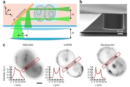
Schematic showing the soSPIM technique. A) Cells can be grown in the microfabricated wells, holding them in place for imaging. B) The 45o micromirrors reflect the excitation beam (dotted line) from a single objective, through the sample, and the resultant emitted fluorescence signal is captured by the same objective.
The soSPIM technique, developed by Professor Virgile Viasnoff, principal investigator at MBI, Singapore, and CNRS, France, utilizes an array of micromirrored wells, where each mirror is inclined at precisely 45 degrees. Together with a beam steering add-on unit, these micromirrors allow for both the excitation beam and resulting fluorescence signal to pass through a single standard, objective lens. The soSPIM technique exhibited fast response and good sectioning capability for 3D imaging of a whole cell up to 30 micrometres above the coverslip and could identify single proteins, deep within the cell. When larger fabricated mirrors were introduced, the system was capable of imaging whole Drosophila embryos.
"Bearing in mind that a typical human skin cell is 30 micrometres in height, most super-resolution microscopy techniques can only capture 2D images near the surface of the glass coverslip or 3D images within the first micrometre," says Prof. Viasnoff. "Although SPIM enables 3D super-resolution imaging of thicker samples at a single-cell level by selectively illuminating a single plane of the specimen with a focused light sheet from one side while capturing the fluorescence signal through a second objective positioned perpendicularly to the light sheet, this approach requires a complicated 2-objective system and special sample holder. This is incompatible with standard microscope systems and prohibitively complicated and expensive for most labs."
"Our soSPIM method is versatile enough to identify single proteins anywhere within a cell or assess cellular organization in whole embryos in 3D on a microscope likely to be found in the majority of life science labs. What's more, we found that soSPIM's acquisition rate was limited solely by sample brightness and the camera's maximum acquisition rate. In this regard, we used an Andor Neo 5.5 sCMOS camera that had proved reliable in the past. Its large, 22mm diameter, 5.5 megapixel sensor is ideal for cell microscopy, and the frame rate of 30 fps sustained or 100 fps burst mode coped easily with our work."
According to Dr. Orla Hanrahan, Imaging Application Specialist at Andor Technology, an Oxford Instruments company:
Professor Viasnoff and colleagues have signalled that it is possible to dramatically simplify the implementation of light sheet microscopy down to the molecular level.
Also, since the microfabricated mirror and sample holder is produced independently of the microscope system, soSPIM is compatible with standard inverted microscopes and high numerical aperture immersion objective lenses. This should allow many more laboratories to study the activity of single proteins within individual cells or tissues, providing new insight into protein function and accelerating our understanding of how protein dysregulation leads to disease. Their innovative approach took advantage of the large field of view, high sensitivity, and fast frame rate capabilities of Andor's Neo sCMOS detector and unleashes a host of possibilities for the future of imaging.
This is not the first time that Andor's sCMOS camera has been at the heart of advances in light sheet microscopy. Both Jan Huisken and his research team from the Max Plank Institute in Dresden, Germany and Philipp J Keller and his research group from the Howard Hughes Medical Institute in Virginia, USA, relied on it for their ground-breaking light sheet research.
 Tell Your Story Even Better with Imaris