An interview with Phil Salmon from Bruker, discussing the development of the SKYSCAN 1276, an in vivo micro-CT scanner designed for preclinical applications.
Please can you give a brief introduction to your work at Bruker?
I work in Bruker micro-CT as a life science applications scientist. Essentially, my role is to serve as an interface between the community of users and the designers, engineers and programmers that make our products.
It's an exciting process sharing new technologies with the researchers who are at the leading edge of their respective fields and able to really put the technologies into practice and realize important scientific advances in partnership with us.
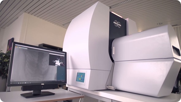
SkyScan 1276
SkyScan 1276 from AZoNetwork on Vimeo.
Can you please give an overview of the new SKYSCAN 1276 that has recently been introduced?
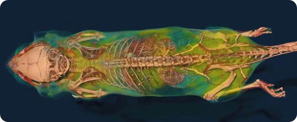
The SKYSCAN 1276 is an in vivo micro-CT scanner, which means it can scan a mouse, rat, or other preclinical model live, while the animal is under anesthesia. That conveys important advantages to life science research: what we call longitudinal scanning.
The same animal can be scanned at different time points, separated by several weeks or months and we can see processes in the animal developing over time.
In vivo scanning has been around since about the turn of the millennium. We were one of the first companies to bring it to the market in 2002 and we have had experience in all the years since in constantly upgrading and developing that technology, which brings us to the release of this very important new scanner, the SKYSCAN 1276.
How does the SKYSCAN 1276 differ from previous technologies?
The SKYSCAN 1276 brings an unprecedented level of imaging performance to in vivo micro-CT. The shortest scan time, for example, is 3.9 seconds and the smallest pixel size is 2.8 microns.
Equally as important is the very high resolution and the new ability to adjust the magnification continually and smoothly between 2.8 microns and larger pixel sizes and larger fields of view, rather than having to choose between a small number of fixed magnifications or only a single magnification and field of view.
This is important because it gives the researcher the flexibility to choose exactly the right balance of resolution, pixel size and field of view, depending on the part of the animal they want to image and also the size of the specimen that they are imaging.
What advantages does the 1276 bring to the preclinical imaging community?
The SKYSCAN 1276 brings technologies which have been pioneered in hospital CT scanning and makes them available to pre-clinical micro-CT in vivo imaging. Foremost among those is helical scanning, where the scan acquisition advances continuously with the rotation of the source and camera.
This confers important advantages; it's artifact free, it's continuous and it improves the quality of the image that you get, for example, of a whole mouse.
Gated imaging or synchronized imaging is an important aspect of micro-CT scanning in vivo and the SKYSCAN 1276 enables a step forward in the performance of gating imaging on the basis of faster image output from the camera.
This means that the synchronized images that are obtained from the breathing cycle and the cardiac cycle, where multiple images per rotation step are combined with monitoring information about breathing or about the heartbeat, are better quality and are brought to a new level of both spatial and temporal resolution.
The X-ray camera is at the heart of any micro-CT imaging system, including in vivo and the X-ray camera of the SKYSCAN 1276 has a pixel format of 11 megapixels, which means four thousand pixels across the image field of view. This can be extended to 8,000 pixels by offset imaging.
This very large pixel format gives the user unprecedented flexibility in terms of combining the ideal size of field of view with the resolution.
Who will benefit from the introduction of this new product?
The SKYSCAN 1276 scanner will bring important advantages to the established users of micro-CT in vivo, such as the bone research community and lung researchers.
However, it will also benefit a much wider range of healthcare fields. For example, the ability to scan a whole mouse with a helical spiral scan will improve the quality of body composition data such as fat fraction and lean tissue volume. This is important in the study of obesity, diabetes and metabolism.
The high resolutions that are possible and the 2.8-micron pixel size will be very valuable in the bone field, in the study of morphometry of mouse and rat trabecular bone, for example.
The very fast scans that are possible and the 3.9 second scan cycle will open up possibilities in biokinetics research, such as the clearance of contrast agents through the kidney.