An interview with Dr. Ichio Aoki, National Institutes for Quantum and Radiological Science and Technology.
Please can you give a brief introduction to yourself and to your role here at the National Institutes of Radiological Science (NIRS) at the Quantum and Radiological Science and Technology (QST)?
I'm an MR imaging scientist in the preclinical field. I'm a team leader of the functional and molecular imaging team at the National Institute of Radiological Sciences (NIRS) in QST.
QST National Institutes for Quantum and Radiological Science and Technology is a new organization, which was launched in April 2016. QST has an imaging research section as well as a synchrotron, and nuclear fusion and high-power laser development sections.
My role at QST is to develop new magnetic resonance imaging technologies and contrast agents in the preclinical field.
Advanced Anatomical and Functional MRI
Advanced Anatomical and Functional MRI from AZoNetwork on Vimeo.
Can you give a brief overview of the research that you and your group do at NIRS?
The predominant theme of our research is to develop new in vivo imaging technologies that can recognize disease, evaluate the benefits of therapies, and discover new diagnostic biomarkers for preclinical applications.
We are working to develop advanced anatomical and functional MRI to reveal the brain function, and to develop functional contrast agents and nanoprobes that can visualize and characterize brain or tumor function, microstructure, and pathologies.
We are focusing on three research fields:
- We are interested in small molecular functional contrast agents and preclinical applications such as manganese-enhanced MRI, nitroxyl radicals as a redox mapping, and ex vivo micro-imaging with contrast agents.
- We are developing nanoparticle-based contrast agents in-house and with some collaborators such as nanomicelles, PEGylated liposomes, iron-oxide nanoparticles, nanogels, and quantum dots. Nanomicelle is one of the most important approaches. We are collaborating with Prof. Kataoka at the Kawasaki Nanomedicine Innovation Center and the University of Tokyo. Nanoparticles have many advantages for multimodal imaging, designing of responsive agents, and diagnostic approaches.
- We are also trying to improve functional MRI methodology with established functional MRI and resting-state functional MRI for mice, rats, marmosets, and macaques. Particularly, we are trying to improve resting-state functional MRI on marmosets using 7Tesla MRI in collaboration with Dr. Afonso C. Silva from NIH and as part of the Brain/MINDS Project with RIKEN and the University of Tokyo, we are developing better functional MRI and microstructure imaging for marmosets. Multiscale and multiparametric understanding using functional and anatomical MRI are the key for the future.
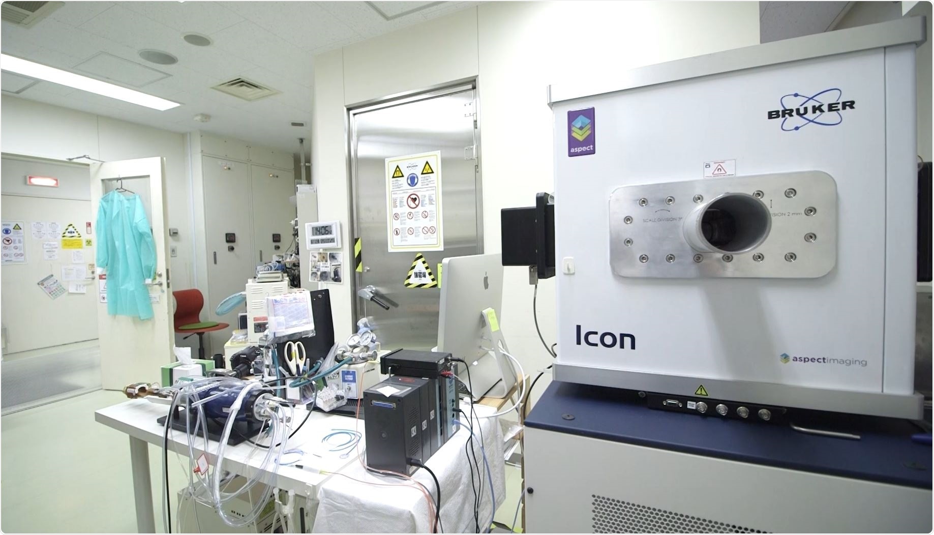
Why did you decide to acquire the 1Tesla ICON system?
The reason for purchasing ICON is simple: that is for the development of contrast agents and the preclinical applications.
Almost seven years ago, we developed a nano-micelle-based gadolinium contrast agent with Prof. Kataoka. Although the contrast agent had excellent T1 reactivity at low-fields, it lost the contrast capability at 7Tesla.
Most of nanoparticle-based contrast agents have both high T1 and T2 reactivity. It means that low-field MRI can have better contrast. Therefore, we considered the 1Tesla MRI system.
There are two reasons for choosing the ICON system at NIRS. First, compatibility with the 7Tesla system. We have some data servers for preclinical MRI. The compatibility of the ICON data is important for us.
The second reason is to do with accuracy. The resonance frequency as a permanent magnet depends on the room temperature. ICON with ParaVision software can perform a frequency correction automatically.
What are the advantages of using the 1Tesla ICON system?
In addition, quantitative mapping requires very high accuracy of measurements such as exact 180-degree RF power, special homogeneity, and sharp slicing. I confirmed that the ParaVision software has high accuracy, with anatomical imaging as well as with quantitative mapping.
High-field MRI has great signal-to-noise ratio, therefore, it has many advantages for micro-imaging, BOLD-based functional MRI, MR spectroscopy, and multinuclear imaging. Especially, 7Tesla MRI can provide unbelievable SNR for mouse brain imaging and tumor microstructure.
Nanoparticles in MRI For Improving Contrast Agents
Nanoparticles in MRI For Improving Contrast Agents from AZoNetwork on Vimeo.
On the other hand, 1Tesla ICON system can provide high-contrast imaging with contrast agents and it is very easy to use. It is also important that the contrast is similar to that of clinical MRI scanners.
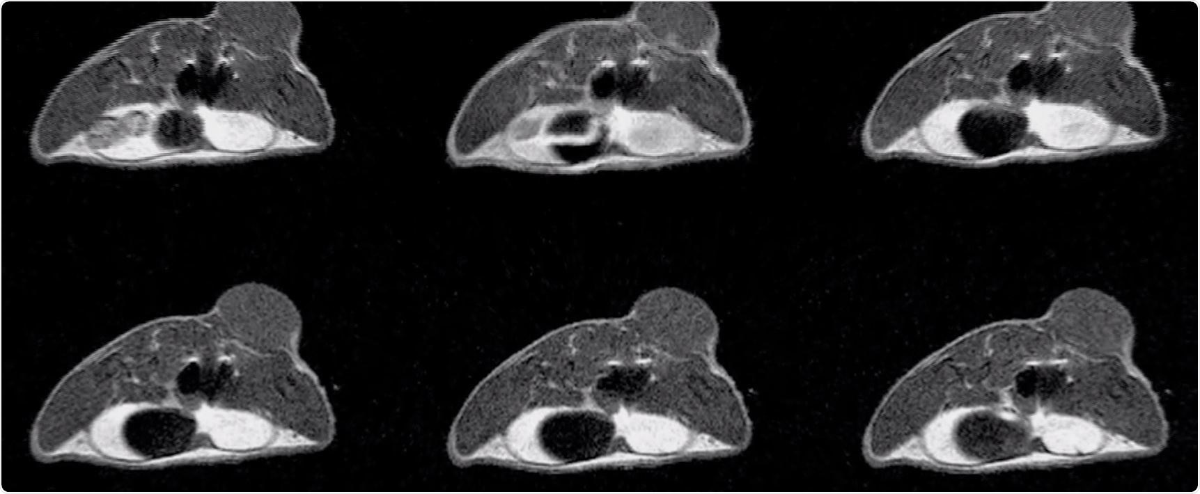
For example, with our manganese with calcium phosphate micelle, we used 7Tesla system for MR angiography and MR spectroscopy and ICON system for T1-weighted MRI. I believe that it is the optimal combination for the application.
What are your plans for your future research?
I always discuss with many researchers and recommend optimal systems for their research purposes. Also, I talk with MR technologists Nitta-san and Ozawa-san and animal technologists Shibata-san and Hayashi-san for improvement of our research environment. I'd like to continue the study of functional and responsive contrast agent development such as manganese, nitroxyl radicals, and diagnostic agents using micelle, nanogels, and liposomes.
In addition, I have two plans for the near future. First, development of safer contrast agents. We believe that we can provide good contrast with the use of gadolinium.
Secondly, we call it a companion nano-imaging. Many kinds of nanomedicine have great advantages, particularly to reduce side effects. However, it is known that the drug accumulation into the focus have big dispersion. I believe that nanoparticle-based contrast agents can act as a predictive marker for nanomedicine, this will allow us to estimate the therapeutic effect before administration.
We will launch an alliance soon for next-generation MR imaging and contrast agent development. I hope that the alliance will provide an open innovation platform in Japan forboth academia and industry.
About Dr. Ichio Aoki - Ph.D 
Team leader at the National Institute of Radiological Sciences (NIRS) April 2007 – Present
Senior Scientist at the NIRS October 2006 – March 2007.
Before that he was a visiting scientist and visiting associate at the NIRS between December 2005 and September 2006.
He spent 2 years as an Assistant Professor at the Department of Medical Informatics at the Meiji University of Oriental Medicine from April 2004 to March 2006.
Has been a visiting Fellow and special volunteer at the National Institutes of Health from April 2000 to July 2003.
 International approaches to tackling food fraud