Logos Biosystems has released the all-new CELENA® X integrated High Content Imaging System, providing the complete solution for rapid high content image acquisition and quantitative analysis.
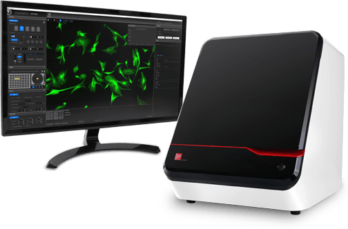
The new CELENA® X produces powerful digital imaging in four modes, while laser autofocusing and motorized positioning of the XYZ stage ensures rapid, reproducible and clear images every time. Its capabilities extend from the simplest fixed cell assays to more complicated, time-lapse live cell assays, making it ideal for high content analysis.
The intuitive CELENA® X Explorer user interface makes creating imaging protocols so straightforward that scientists with limited imaging experience will have no problem setting up high content imaging experiments. Logos Biosystems is offering the CELENA® X system at a very affordable base cost to increase accessibility to high content imaging experiments.
We are excited about introducing the CELENA® X High Content Imaging System to the market. It starts at an affordable price point within the reach of most individual labs’ budgets with the option to upgrade for higher levels of performance.”
Ana Kim, US Regional Director, Logos Biosystems.
The CELENA® X is compatible with Olympus and Zeiss objectives, has interchangeable hard-coated LED fluorescence filters and the facility for on-stage incubation, allowing precise physiological and non-physiological conditions for a range of live cell imaging applications.
Seamless integration of imaging and data analysis processes
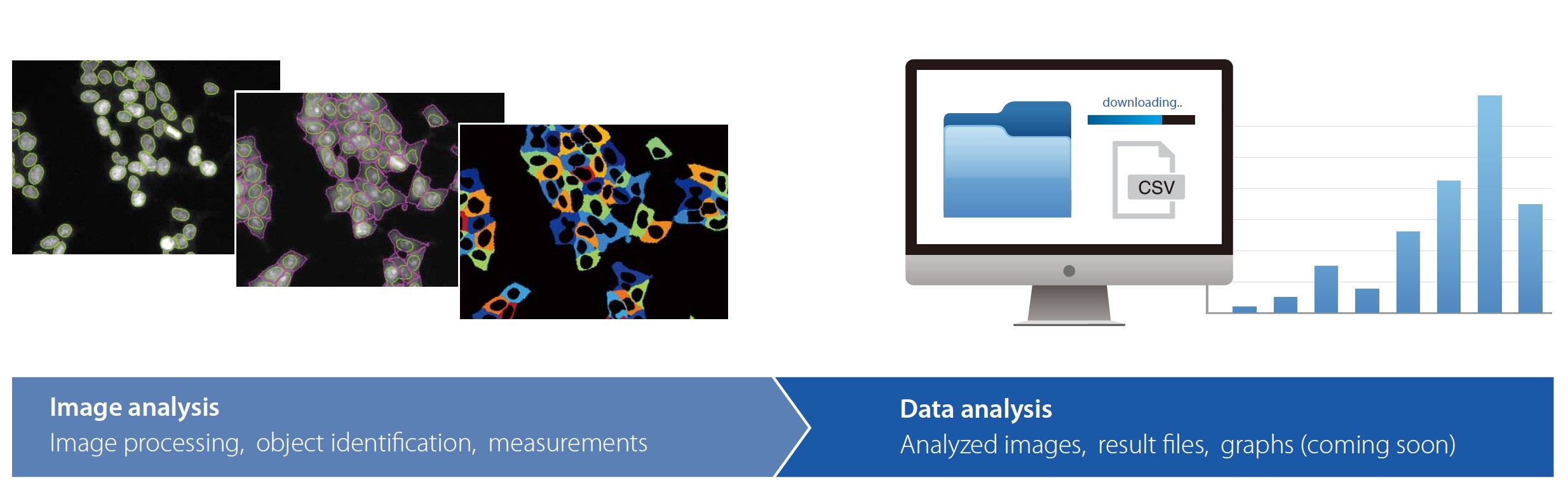
The CELENA® X comes with built-in Cell Analyzer software, that processes the acquired images and data for quantitative analysis, producing statistically robust data for a variety of experimental methodologies. The Cell Analyzer software can also be readily customized to create personalized workflows for high content analysis.
Customizable High Content Analysis. Create and customize image analysis projects. Quantitatively analyze multiple image-based phenotypes
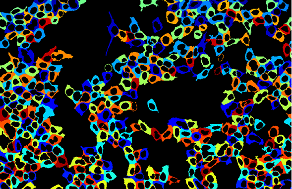
Analysis pipelines can be developed and reused to identify cellular or sub-cellular objects, process images for optimal data collection, and make various measurements. Image analysis can involve differentiation of multiple phenotypes, determination of the morphology of individual cells and organelles and defining the spatial distribution of targets. Multiple measurements can be made for each cell.
Image analysis can be conducted directly on the CELENA® X or remotely by downloading CELENA® X Cell Analyzer onto a personal computer.
CELENA® X Applications
The CELENA® X is as flexible as it is powerful and can be employed for a vast array of investigations into cell function, including apoptosis, autophagy, proliferation, and migration, as well as studies of cytotoxicity, cell viability and transfection efficiency.
With the power to capture and quantify cellular information in both fixed and live cells, the CELENA® X provides a valuable tool for life science research as well as drug discovery and development. The CELENA® X can be used to determine cell distribution, image histopathological biopsies, quantify transfection efficiency and evaluate the cytotoxic effects of drugs among other applications.
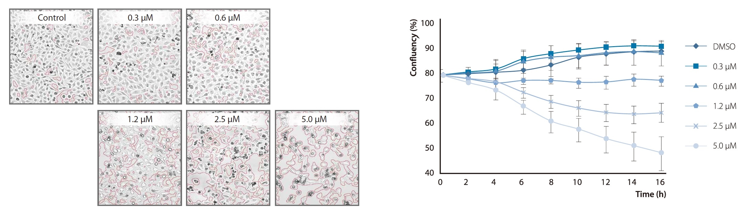
Evaluating the cytotoxic effects of Camptothecin on HeLa cells with the CELENA® X.
Confluent HeLa cells were treated with varying doses of Camptothecin (CPT) at 2 hour intervals for 6 hours and subsequently imaged to determine how CPT concentration affects cell viability. CELENA®X Cell Analyzer was used to measure the confluency of cells in a multi-well plate in a rapid, reproducible, and quantifiable manner