What are the three modes of neurotransmission, and how do they differ in terms of temporal coupling with action potentials?
There are three modes of neurotransmission: stimulation-dependent release (evoked), asynchronous evoked release, and spontaneous release.
The stimulation-dependent release is tightly connected with incoming action potentials, which means that when an action potential is received, synaptic vesicles fuse with the presynaptic membrane practically instantly, releasing neurotransmitters. It is like runners starting as soon as the start gun goes off; the motion is instantaneous and synchronized.
Asynchronous evoked release occurs in response to an action potential, albeit with a delay. It is less temporally related to action potential, similar to runners who did not get off to a fast start. This form of release has a more flexible time.
In contrast, spontaneous release is fully independent of action potentials. Even without stimulation, synaptic vesicles can fuse with the membrane and release neurotransmitters. This is a background process that occurs in the absence of any immediate stimulus from the neuron's electrical activity.
How did the work of Fatts and Katz in the 1950s contribute to our understanding of neurotransmission, particularly regarding spontaneous and evoked release?
Fatts and Katz's work in the 1950s was foundational. They investigated the neuromuscular junction, which connects motor neurons with muscle cells, and utilized microelectrodes to assess electrical activity in these muscle cells.
They discovered that activating the motor neuron resulted in a significant depolarization of the muscle - an evoked response, which is a classic example of action potential-dependent neurotransmission. The exciting element was that scientists identified modest, spontaneous electrical potentials even when the cell was not activated.
These modest, random events demonstrated that neurotransmitter release can occur without an action potential. This discovery set the framework for our current understanding of spontaneous and evoked neurotransmission.
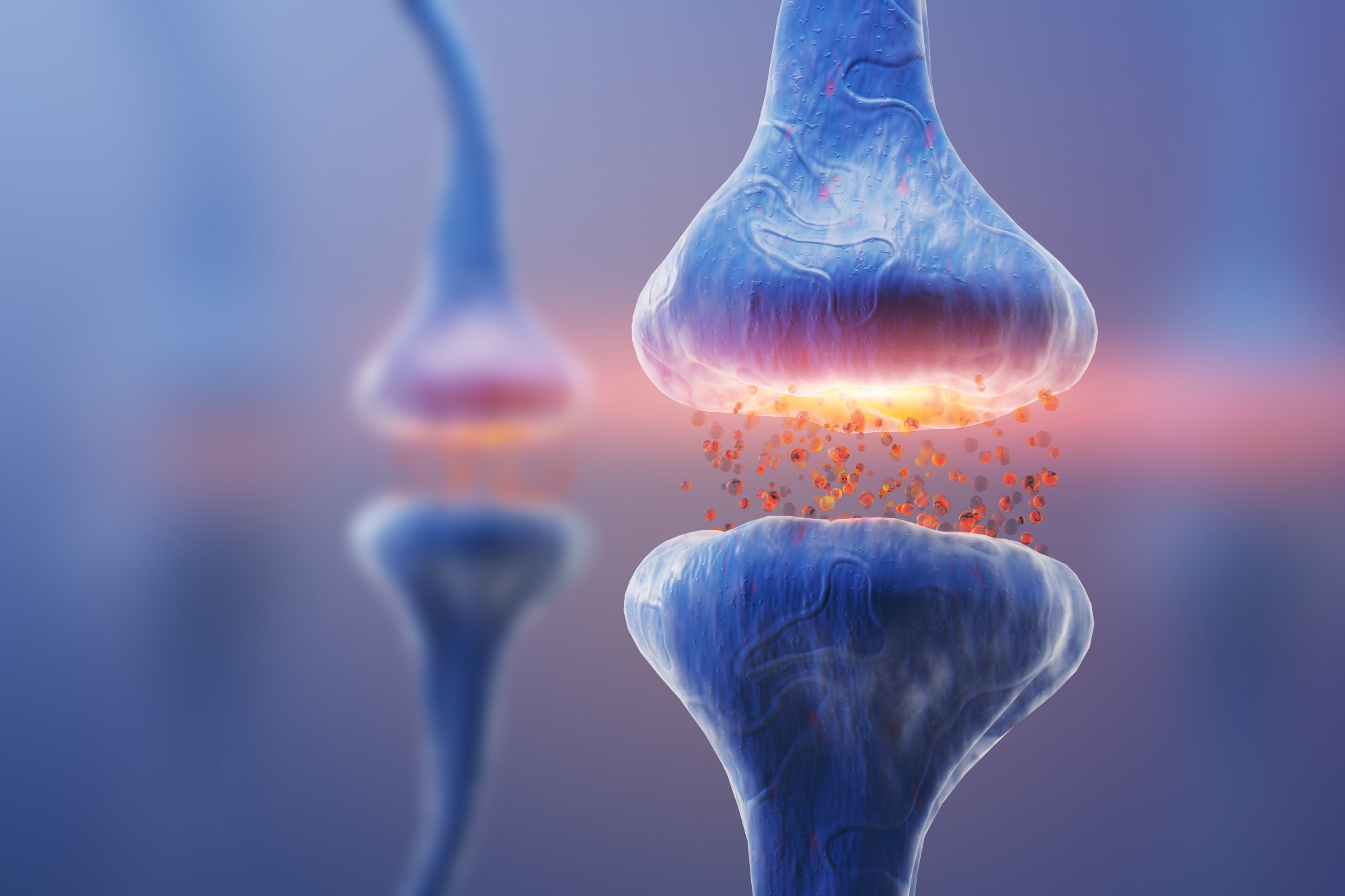
Image Credit: KateStudio/Shutterstock.com
What role does gephyrin play in GABAergic synapses, and how do Artemisinins affect the gephyrin scaffold structure?
Gephyrin is important in GABAergic synapses because it serves as a scaffolding protein. It is in charge of anchoring GABA-A receptors in the synapse, ensuring that they are appropriately positioned to facilitate inhibitory neurotransmission. Without gephyrin, these receptors would not cluster efficiently, causing synaptic dysfunction.
When we treat neurons with anti-malarial drugs called artemisinins, we disrupt this scaffolding structure. Artemisinins bind to the gephyrin universal binding pocket, which also hosts GABA-A receptors. As a result, the volume of gephyrin clusters decreases, indicating that the scaffolding diminishes.
This shrinking is limited to gephyrin at the synapse, leaving the presynaptic markers unchanged. This disturbance lowers the synapse's ability to anchor GABA-A receptors, which has important functional consequences for neurotransmission.
How does the segregation of neurotransmission occur within the same synapse, and what evidence supports this phenomenon in both evoked and spontaneous release?
Neurotransmission is segregated within the same synapse because different parts of the synapse preferentially support provoked or spontaneous release.
At a single synapse, the presynaptic and postsynaptic machinery involved in the evoked release is more densely concentrated in some locations, whereas other areas may be more specialized for spontaneous release.
Evidence for this comes from trials in which we utilized MK-801, a use-dependent NMDA receptor blocker, to differentiate between these modes. For example, it was discovered that different NMDA receptor populations are active during spontaneous neurotransmission vs provoked release.
In addition, we have observed structural differences in presynaptic and postsynaptic protein density across regions that support these various forms of release. In other words, even inside the same active zone of a synapse, we have specialized structures that enable these two forms of neurotransmission.
What impact does the disruption of gephyrin by Artemisinin treatment have on spontaneous inhibitory neurotransmission and tonic inhibition?
Artemisinin medication largely influences spontaneous inhibitory neurotransmission. After only one hour of Artemisinin therapy, we find a considerable drop in the frequency of micro inhibitory postsynaptic currents (mini IPSCs), which indicates spontaneous neurotransmission.
This occurs when gephyrin's scaffolding is disturbed, preventing GABA-A receptors from correctly anchoring at the synapse. As a result, even if a synaptic vesicle emits neurotransmitters, there may be insufficient receptors to respond to the signal.
Interestingly, tonic inhibition, which is mediated by extrasynaptic GABA-A receptors, is slightly enhanced. We believe this is because GABA-A receptors that are no longer attached at the synapse might migrate into the extrasynaptic region, boosting tonic inhibition.
How does spontaneous neurotransmission contribute to the regulation of homeostatic plasticity, and what is its potential relevance to neuropsychiatric illnesses?
Homeostatic plasticity, the brain's method of preserving equilibrium in neural circuits, relies heavily on spontaneous neurotransmission. It contributes to the "equilibrium" of synaptic activity, ensuring that neurons do not become excessively excitable or quiescent.
There is an increasing amount of data relating spontaneous neurotransmission to the etiology and treatment of neuropsychiatric conditions.
For example, when medicines like ketamine inhibit NMDA receptors involved in spontaneous transmission, synaptic upscaling occurs, which is a compensatory mechanism in which synapses grow in response to reduced activity.
This phenomenon is associated with ketamine's antidepressant characteristics, implying that modulating spontaneous neurotransmission may provide new therapeutic methods for disorders such as depression.
What were the electrophysiological effects observed after Artemisinin treatment, and how did they differ between spontaneous and evoked neurotransmission?
Artemisinin administration resulted in a selective decrease in spontaneous inhibitory neurotransmission, but evoked neurotransmission remained mostly unaltered. Specifically, the number of micro IPSCs (spontaneous currents) was dramatically reduced. The magnitude of these spontaneous currents also decreased, implying that fewer GABA-A receptors were available to facilitate this sort of transmission.
In contrast, when we examined evoked neurotransmission using repetitive stimulation protocols, we found no change in the amplitude of evoked IPSCs (inhibitory postsynaptic currents) or the paired-pulse ratio, indicating that the mechanisms for action potential-dependent release remained intact. This shows that spontaneous and induced types of neurotransmission can be regulated differently within the same synapse.
How does the loss of gephyrin at the synaptic periphery correlate with changes in GABA receptor clustering and synaptic signaling?
The absence of gephyrin at the periphery of synaptic clusters has a direct effect on GABA-A receptor clustering and synaptic signaling. Artemisinin treatment causes a selective reduction in gephyrin volume at the periphery, leaving the core of the gephyrin cluster relatively intact. As a result, GABA-A receptors anchored in these peripheral locations are eliminated, lowering the total surface area available for inhibitory transmission.
As the number of receptors available to respond to synaptic vesicle release decreases, so does the frequency and amplitude of spontaneous neurotransmission. In short, gephyrin disruption impairs the synaptic scaffold, reducing synaptic communication efficiency, particularly in spontaneous neurotransmission.
Watch the full webinar
About the Speakers

Dr. Kavalali is a Professor & Chair of the Department of Pharmacology and William Stokes Chair in Experimental Therapeutics at Vanderbilt School of Medicine. Dr. Kavalali studies mechanisms of neurotransmission and synaptic signaling in the central nervous system using electrical and optical recording techniques as well as molecular tools. His group focuses on the molecular basis and functional consequences of heterogeneity among synaptic vesicle recycling pathways present within individual synapses.

Natalie is a Ph.D., neuroscience student at Vanderbilt University and a member of the Kavalali Lab. Her current research includes using STORM and electrophysiology recording methods to study synaptic function. Prior to Vanderbilt, she was a Masters Student Research Scientist at University College London.
About Bruker Nano Surfaces and Metrology

Bruker’s suite of fluorescence microscopy systems provides a full range of solutions for life science researchers. Their multiphoton imaging systems provide the imaging depth, speed and resolution required for intravital imaging applications, and their confocal systems enable cell biologists to study function and structure using live-cell imaging at speeds and durations previously not possible. Bruker’s super-resolution microscopes are setting new standards with quantitative single molecule localization that allows for the direct investigation of the molecular positions and distribution of proteins within the cellular environment. And their Luxendo light-sheet microscopes, are revolutionizing long-term studies in developmental biology and investigation of dynamic processes in cell culture and small animal models.
 Bruker announces acquisition of Inscopix for neuroscience research