Technology innovators in super-resolution microscopy, Amsterdam-based Confocal.nl,reports on the implementation of a new turnkey confocal imaging system in the laboratory of Professor Markus Sauer at the University of Würzburg.
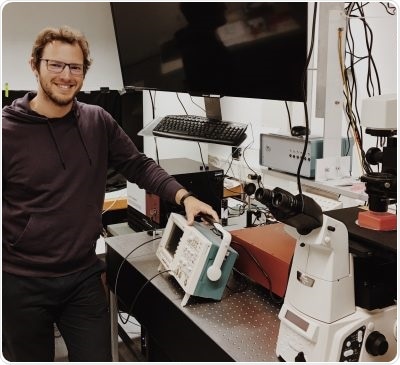
Andreas Kurz with the RCM confocal/dSTORM system located in Professor Markus Sauer’s laboratory at the University of Würzburg
Professor Markus Saueris the Chair of the group for biotechnology and biophysics at the University of Würzburg. He is very interested in imaging: “Imaging technologies are central platforms that drive fundamental research in virtually all disciplines across the biological and medical sciences. By seeing how a system looks, functions can be understood and comparison made between healthy and unhealthy systems such as cells, tissue, a whole organism or population, plants, animals and humans.”
Professor Sauer’s PhD student, Andreas Kurz, describes how he implemented a new technique called Re-scan Confocal Microscopy (RCM):
We integrated the RCM module from Confocal.nl to our existing single molecule dSTORM setup to establish a new correlative imaging technique. As RCM is based on image scanning, it intrinsically has super-resolution capabilities (170 nm) combined with the advantages of a conventional confocal microscope. Now with this combined correlative imaging setup, a new variety of experiments are possible. Recording dynamic processes and the long-term observation of cell activity followed by resolving the structure of interest, a superb resolution of around 20 nm will now be possible.”
Mr Kurz continues to describe the process to select RCM:
Confocal microscopy is an imaging technique with lots of advantages. First of all, it is an easy-to-use tool for routine work in the field of fluorescence. Now with RCM, we have an enhanced version of confocal microscopy which allows the observation of sensitive samples and more complex imaging procedures at a higher resolution than other confocal microscopies. This system is now used to complement other techniques such as structured illumination microscopy, single molecule localization microscopy setups and light sheet illumination microscopy.
We ordered our RCM unit from Confocal.nl. Their CTO, Erik Manders, visited to personally assist with the installation and training. Since then, we have had excellent support with updates through software plugins and exchanging applications experiences. Concerning the setup, we had the microscope body already installed (Nikon Eclipse TiE). In addition, we purchased a Skyra multi-line laser unit from Cobolt and an Andor Zyla 4.2 sCMOS camera. It was great to be able to use our existing microscope and then add specific components to, in effect, produce a turnkey set-up to match our demanding needs. Confocal.nl continue to provide a lot of support (local as well as via mail/phone). Having an accessible and manageable team, we get the ease of communication with a personal touch, something I really prefer in this environment.”
Concluding, Mr Kurz says:
Now, our experimental set-up is a unique, fast-scanning, resolution-enhanced and live-cell capable system configured for both dSTORM and RCM that delivers super-resolution performance. Our main reasons for purchase have been realized in terms of performance of the complete package integrating our existing components at a very fair price. We have found the multi-color capability with incredibly low laser power very useful for live cell imaging.”
Speaking about the installation at Würzburg, Confocal.nl’s CEO, Peter Drent, says;
We were extremely pleased to have Professor Sauer’s team select the RCM. They provided precise requirements from which we have been able to configure an excellent, working system with high resolution and sensitivity. While dSTORM is quite an intensive experimental technique involving complex biological and chemical sample preparation, it does provide 20 nm resolution. This is now complemented by RCM also giving super-resolution at 170 nm with minimal sample preparation delivering a simple, instant image.”